The Rheumatologist
COVID-19 News
  • Connect with us:
  • Facebook
  • Twitter
  • LinkedIn
  • YouTube
  • Feed
  • Home
  • Conditions
    • Rheumatoid Arthritis
    • SLE (Lupus)
    • Crystal Arthritis
      • Gout Resource Center
    • Spondyloarthritis
    • Osteoarthritis
    • Soft Tissue Pain
    • Scleroderma
    • Vasculitis
    • Systemic Inflammatory Syndromes
    • Guidelines
  • Resource Centers
    • Axial Spondyloarthritis Resource Center
    • Gout Resource Center
    • Psoriatic Arthritis Resource Center
    • Rheumatoid Arthritis Resource Center
    • Systemic Lupus Erythematosus Resource Center
  • Drug Updates
    • Biologics & Biosimilars
    • DMARDs & Immunosuppressives
    • Topical Drugs
    • Analgesics
    • Safety
    • Pharma Co. News
  • Professional Topics
    • Ethics
    • Legal
    • Legislation & Advocacy
    • Career Development
      • Certification
      • Education & Training
    • Awards
    • Profiles
    • President’s Perspective
    • Rheuminations
    • Interprofessional Perspective
  • Practice Management
    • Billing/Coding
    • Quality Assurance/Improvement
    • Workforce
    • Facility
    • Patient Perspective
    • Electronic Health Records
    • Apps
    • Information Technology
    • From the College
    • Multimedia
      • Audio
      • Video
  • Resources
    • Issue Archives
    • ACR Convergence
      • Systemic Lupus Erythematosus Resource Center
      • Rheumatoid Arthritis Resource Center
      • Gout Resource Center
      • Abstracts
      • Meeting Reports
      • ACR Convergence Home
    • American College of Rheumatology
    • ACR ExamRheum
    • Research Reviews
    • ACR Journals
      • Arthritis & Rheumatology
      • Arthritis Care & Research
      • ACR Open Rheumatology
    • Rheumatology Image Library
    • Treatment Guidelines
    • Rheumatology Research Foundation
    • Events
  • About Us
    • Mission/Vision
    • Meet the Authors
    • Meet the Editors
    • Contribute to The Rheumatologist
    • Subscription
    • Contact
  • Advertise
  • Search
You are here: Home / Articles / Case Report: Rapidly Progressive Interstitial Lung Disease in a 6-Year-Old

Case Report: Rapidly Progressive Interstitial Lung Disease in a 6-Year-Old

April 17, 2021 • By Anusha Vuppala, MD, & Sarwat Umer, MD

  • Tweet
  • Email
Print-Friendly Version / Save PDF
The patient presented with heliotrope rash.

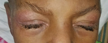
Photo 1: The patient presented with heliotrope rash.

Clinically amyopathic dermatomyositis (CADM), a rare subset of dermatomyositis (DM), is an autoimmune disease characterized by cutaneous findings of typical DM without evidence of myositis. Childhood presentation of CADM is rare, and not many studies describe the epidemiology of juvenile CADM.1,2

You Might Also Like
  • Case Report: Interstitial Lung Disease with Positive ANCA Test
  • Fellows’ Forum Case Report: Progressive Weakness and Debilitation with Skin Rash
  • Cyclophosphamide for Connective Tissue Disease-Associated Interstitial Lung Disease
Explore This Issue
April 2021
Also By This Author
  • Case Report: Digital Ulcers & Weight Loss in a Pediatric Patient

Although lung disease is rare among patients with juvenile DM, a few reports have been published since 2007 about interstitial lung disease (ILD) in children with CADM.3 Rapidly progressive ILD occurs more in CADM patients than in classic DM patients and has a poor prognosis.

ad goes here:advert-1
ADVERTISEMENT
SCROLL TO CONTINUE

Several studies have been conducted to find the association between specific auto­antibodies and ILD in patients with CADM. Autoantibodies to a 140 kDa cytoplasmic protein (i.e., anti-CADM-140 antibodies) were first described in adults with CADM in Japan.4,5 The antibody is directed against an RNA helicase encoded by melanoma differentiation-associated gene 5 (MDA5), which is a pattern recognition receptor that can induce the transcription of type I interferon genes in response to viral infection.6

Later, a strong association was seen between rapidly progressive ILD and the presence of anti-CADM-140 antibodies, also known as anti-MDA5 antibodies, in juvenile patients with both classic DM and CADM.7 Patients with juvenile DM who have anti-MDA5 antibodies can develop rapidly progressive ILD, which is associated with a worse prognosis. Prompt and aggressive inter­vention may improve outcomes, but can sometimes be challenging due to difficulty recognizing the disease, in part because of heterogeneous presentations, and failure to respond to conventional immunosuppression.

ad goes here:advert-2
ADVERTISEMENT
SCROLL TO CONTINUE

We describe a 6-year-old patient who presented with Gottron papules, heliotrope rash, mild muscle weakness and persistent fevers.

Case Description

The patient is a 6-year-old Black child who presented with symptoms of mild muscle weakness and persistent fever up to 103ºF. The febrile episodes occurred almost every other day for about four to five months. He later developed joint pain, mainly involving his knees and elbows, with erythema and swelling. His mother noted a rash around her son’s eyes and on his hands about a month prior to presentation.

The patient was seen by his primary care physician and laboratory tests were obtained (see Table 1). The patient was referred to our hospital for further evaluation.

ad goes here:advert-3
ADVERTISEMENT
SCROLL TO CONTINUE

On examination, the patient was noted to have a heliotrope rash (see Photo 1) and Gottron papules (see Photo 2), Raynaud’s phenomenon and digital ulcers on the distal tips of his thumb, index and middle finger (see Photo 3); and mild weakness of his arms and legs.

Magnetic resonance imaging of both legs showed a mild increase in signal intensity in the rectus femoris, vastus lateralis, vastus intermedius and hamstring muscles in both legs. Treatment was initiated with 30 mg/kg/day intravenous steroids for three days and 10 mg of sub­cutaneous methotrexate once a week. He experienced an initial improvement of his weakness and rash. However, during the intervening four weeks before his follow-up appointment, the weakness and rash worsened. Steroid administrations were repeated, and 1 gm/kg intravenous immunoglobulin was administered once daily for two days.

Pages: 1 2 3 4 | Single Page

Filed Under: Conditions Tagged With: Clinically Amyotrophic Dermatomyositis (CADM), dermatomyositis, interstitial lung disease (ILD), juvenile dermatomyositis, Pediatric RheumatologyIssue: April 2021

You Might Also Like:
  • Case Report: Interstitial Lung Disease with Positive ANCA Test
  • Fellows’ Forum Case Report: Progressive Weakness and Debilitation with Skin Rash
  • Cyclophosphamide for Connective Tissue Disease-Associated Interstitial Lung Disease
  • Case Report: A Patient with Clinically Amyotrophic Dermatomyositis & Associated ILD & RA Overlap

American College of Rheumatology

Visit the official website for the American College of Rheumatology.

Visit the ACR »

Simple Tasks

Learn more about the ACR’s public awareness campaign and how you can get involved. Help increase visibility of rheumatic diseases and decrease the number of people left untreated.

Visit the Simple Tasks site »

Meeting Abstracts

Browse and search abstracts from the ACR Convergence and ACR/ARP Annual Meetings going back to 2012.

Visit the Abstracts site »

The Rheumatologist newsmagazine reports on issues and trends in the management and treatment of rheumatic diseases. The Rheumatologist reaches 11,500 rheumatologists, internists, orthopedic surgeons, nurse practitioners, physician assistants, nurses, and other healthcare professionals who practice, research, or teach in the field of rheumatology.

About Us / Contact Us / Advertise / Privacy Policy / Terms of Use

  • Connect with us:
  • Facebook
  • Twitter
  • LinkedIn
  • YouTube
  • Feed

Copyright © 2006–2022 American College of Rheumatology. All rights reserved.

ISSN 1931-3268 (print)
ISSN 1931-3209 (online)

loading Cancel
Post was not sent - check your email addresses!
Email check failed, please try again
Sorry, your blog cannot share posts by email.