The Rheumatologist
COVID-19 News
  • Connect with us:
  • Facebook
  • Twitter
  • LinkedIn
  • YouTube
  • Feed
  • Home
  • Conditions
    • Rheumatoid Arthritis
    • SLE (Lupus)
    • Crystal Arthritis
      • Gout Resource Center
    • Spondyloarthritis
    • Osteoarthritis
    • Soft Tissue Pain
    • Scleroderma
    • Vasculitis
    • Systemic Inflammatory Syndromes
    • Guidelines
  • Resource Centers
    • Ankylosing Spondylitis Resource Center
    • Gout Resource Center
    • Rheumatoid Arthritis Resource Center
    • Systemic Lupus Erythematosus Resource Center
  • Drug Updates
    • Biologics & Biosimilars
    • DMARDs & Immunosuppressives
    • Topical Drugs
    • Analgesics
    • Safety
    • Pharma Co. News
  • Professional Topics
    • Ethics
    • Legal
    • Legislation & Advocacy
    • Career Development
      • Certification
      • Education & Training
    • Awards
    • Profiles
    • President’s Perspective
    • Rheuminations
  • Practice Management
    • Billing/Coding
    • Quality Assurance/Improvement
    • Workforce
    • Facility
    • Patient Perspective
    • Electronic Health Records
    • Apps
    • Information Technology
    • From the College
    • Multimedia
      • Audio
      • Video
  • Resources
    • Issue Archives
    • ACR Convergence
      • Systemic Lupus Erythematosus Resource Center
      • Rheumatoid Arthritis Resource Center
      • Gout Resource Center
      • Abstracts
      • Meeting Reports
      • ACR Convergence Home
    • American College of Rheumatology
    • ACR ExamRheum
    • Research Reviews
    • ACR Journals
      • Arthritis & Rheumatology
      • Arthritis Care & Research
      • ACR Open Rheumatology
    • Rheumatology Image Library
    • Treatment Guidelines
    • Rheumatology Research Foundation
    • Events
  • About Us
    • Mission/Vision
    • Meet the Authors
    • Meet the Editors
    • Contribute to The Rheumatologist
    • Subscription
    • Contact
  • Advertise
  • Search
You are here: Home / Articles / Case Study in Dermatology: Familial Sarcoidosis/Blau Syndrome

Case Study in Dermatology: Familial Sarcoidosis/Blau Syndrome

June 1, 2014 • By Johanna Sheu, MS, & Joseph F. Merola, MD, MMSc

  • Tweet
  • Email
Print-Friendly Version / Save PDF

You Might Also Like
  • Case Study in Dermatology: Tender Papules on Elbows, Hands in RA
  • Dermatology Case Review
  • Dermatology Case Answer
Explore This Issue
June 2014
Multiple erythematous, slightly hyperpigmented waxy papules coalescing into plaques without scale over the bilateral distal lower extremities.
Figure 1: Multiple erythematous, slightly hyperpigmented waxy papules coalescing into plaques without scale over the bilateral distal lower extremities.

ad goes here:advert-1
ADVERTISEMENT
SCROLL TO CONTINUE

The Case

A 43-year-old woman presented with a recurrent rash on her legs. She had a 20-year history of a diagnosis of sarcoidosis, which had been complicated by uveitis and inflammatory arthritis. She also had a history of a subacute granulomatous thyroiditis. Multiple family members also had similar episodes of uveitis, diagnoses of inflammatory arthritis and “sarcoid-like” illnesses.

On examination, she had multiple erythematous, slightly hyperpigmented waxy papules coalescing into plaques without scale over both distal lower extremities (see Figure 1).

ad goes here:advert-2
ADVERTISEMENT
SCROLL TO CONTINUE

What’s Your Diagnosis?

  1. Majocchi’s granuloma/tinea infection
  2. Granuloma annulare
  3. Palisaded neutrophilic granulomatous dermatitis
  4. Familial sarcoidosis/Blau syndrome

Correct answer: D

Familial Sarcoidosis/Blau Syndrome

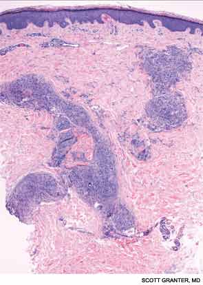
A skin biopsy of a representative lesion was performed, and histopathologic examination revealed nodular aggregates of histiocytes forming non-necrotizing, “naked” granulomas (see Figure 2). Special stains and PCR studies for infectious organisms including mycobacteria were negative. Genetic testing revealed that our patient and multiple family members had a base substitution in the CARD15/NOD2 gene, consistent with a diagnosis of Blau syndrome. Topical steroids were prescribed for her lesions, which resolved with treatment.

Blau syndrome was first described in 1985 by Edward Blau, a pediatrician. It’s a rare, autosomal dominant autoinflammatory disorder with a typical onset within the first two decades of life. Clinically, patients most commonly present with a triad of granulomatous dermatitis, symmetric arthritis and recurrent uveitis. Patients may also have other organ involvement, including hepatic, renal, cardiovascular or neurologic complications.1,2 Mutations that cause Blau syndrome have been mapped to CARD15/NOD2, a caspase recruitment domain gene.

ad goes here:advert-3
ADVERTISEMENT
SCROLL TO CONTINUE

The CARD15/NOD2 gene contains three domains. It is most commonly known for mutations in the third domain, associated with Crohn’s disease, which involves granulomatous inflammation of the bowel. Mutations in the second domain are associated with both Blau syndrome and early onset sarcoidosis. CARD15/NOD2 activates NF-kB in the setting of bacterial cell wall components.3 In Blau syndrome, genetic mutations in the second domain are usually gain-of-function mutations, leading to increased NF-kB activation and possibly increased activity of other inflammatory cytokines.4

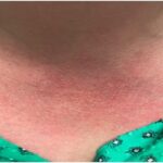
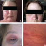
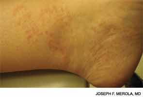
Blau syndrome often presents earliest and most commonly in the skin. Patients present with multiple erythematous and waxy dermal papules and plaques with a lack of scale. Skin biopsy is helpful in diagnosis because it often demonstrates granulomatous inflammation.5

Histopathologic examination revealed nodular aggregates of histiocytes forming non-necrotizing, “naked” granulomas.
Figure 2: Histopathologic examination revealed nodular aggregates of histiocytes forming non-necrotizing, “naked” granulomas.

Discussions of Other Choices

Majocchi’s granuloma occurs when a dermatophyte infection involves the deeper structures of the skin, such as the hair follicles and the dermis. These lesions occur commonly in areas with hair follicles, such as the legs. Patients can also demonstrate other evidence of derma­tophyte infection, such as onychomycosis or tinea pedis. Histopathologic examination of Majocchi’s granuloma would reveal fungal forms on the biopsy.

Pages: 1 2 | Single Page

Filed Under: Conditions, Rheumatoid Arthritis, SLE (Lupus), Vasculitis Tagged With: autoinflammatory, Blau syndrome, Dermatology, Rheumatoid arthritis, rheumatologist, Sarcoidosis, Systemic lupus erythematosus, VasculitisIssue: June 2014

You Might Also Like:
  • Case Study in Dermatology: Tender Papules on Elbows, Hands in RA
  • Dermatology Case Review
  • Dermatology Case Answer
  • Dermatology Case: History of ANCA–Associated Vasculitis, Fever, Rash

American College of Rheumatology

Visit the official website for the American College of Rheumatology.

Visit the ACR »

Meeting Abstracts

Browse and search abstracts from the ACR Convergence and ACR/ARP Annual Meetings going back to 2012.

Visit the Abstracts site »

ACR Convergence

Don’t miss rheumatology’s premier scientific meeting for anyone involved in research or the delivery of rheumatologic care or services.

Visit the ACR Convergence site »

The Rheumatologist newsmagazine reports on issues and trends in the management and treatment of rheumatic diseases. The Rheumatologist reaches 11,500 rheumatologists, internists, orthopedic surgeons, nurse practitioners, physician assistants, nurses, and other healthcare professionals who practice, research, or teach in the field of rheumatology.

About Us / Contact Us / Advertise / Privacy Policy / Terms of Use

  • Connect with us:
  • Facebook
  • Twitter
  • LinkedIn
  • YouTube
  • Feed

Copyright © 2006–2021 American College of Rheumatology. All rights reserved.

ISSN 1931-3268 (print)
ISSN 1931-3209 (online)

loading Cancel
Post was not sent - check your email addresses!
Email check failed, please try again
Sorry, your blog cannot share posts by email.
This site uses cookies: Find out more.