Video: Every Case Tells a Story| Webinar: ACR/CHEST ILD Guidelines in Practice

An official publication of the ACR and the ARP serving rheumatologists and rheumatology professionals

  • Conditions
    • Axial Spondyloarthritis
    • Gout and Crystalline Arthritis
    • Myositis
    • Osteoarthritis and Bone Disorders
    • Pain Syndromes
    • Pediatric Conditions
    • Psoriatic Arthritis
    • Rheumatoid Arthritis
    • Sjögren’s Disease
    • Systemic Lupus Erythematosus
    • Systemic Sclerosis
    • Vasculitis
    • Other Rheumatic Conditions
  • FocusRheum
    • ANCA-Associated Vasculitis
    • Axial Spondyloarthritis
    • Gout
    • Psoriatic Arthritis
    • Rheumatoid Arthritis
    • Systemic Lupus Erythematosus
  • Guidance
    • Clinical Criteria/Guidelines
    • Ethics
    • Legal Updates
    • Legislation & Advocacy
    • Meeting Reports
      • ACR Convergence
      • Other ACR meetings
      • EULAR/Other
    • Research Rheum
  • Drug Updates
    • Analgesics
    • Biologics/DMARDs
  • Practice Support
    • Billing/Coding
    • EMRs
    • Facility
    • Insurance
    • QA/QI
    • Technology
    • Workforce
  • Opinion
    • Patient Perspective
    • Profiles
    • Rheuminations
      • Video
    • Speak Out Rheum
  • Career
    • ACR ExamRheum
    • Awards
    • Career Development
  • ACR
    • ACR Home
    • ACR Convergence
    • ACR Guidelines
    • Journals
      • ACR Open Rheumatology
      • Arthritis & Rheumatology
      • Arthritis Care & Research
    • From the College
    • Events/CME
    • President’s Perspective
  • Search

Dermatology Symptoms Point to Connective Tissue Disorder

Natalie A. Wright, MD, and Joseph F. Merola, MD, MMSc, FAAD, FACR  |  Issue: May 2015  |  May 15, 2015

The patient had flexion contractures of her knees.

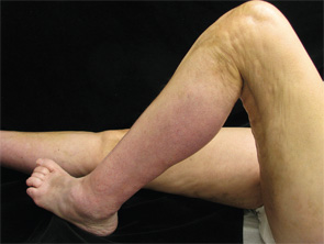
Figure 3. The patient had flexion contractures of her knees.

months from symptom onset was also found to be associated with improved prognosis. A role for immunosuppressive agents in conjunction with corticosteroids has also been discussed in the literature as possibly improving treatment outcomes.2,6 Methotrexate is often the most commonly used corticosteroid-sparing agent; however, the efficacy of mycophenolate mofetil, azathioprine, tumor necrosis factor inhibitors and other therapies has been detailed in small case series. In one retrospective study of 34 patients, methotrexate used in combination with corticosteroids was needed in 44% of patients due to an unsatisfactory response to corticosteroids alone.6

Figure 4. The exam also revealed indurated plaques on the patient's abdomen.

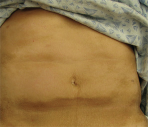
Figure 4. The exam also revealed indurated plaques on the patient’s abdomen.

Because there can be an association with underlying malignancy, most commonly hematologic and myelodysplastic disorders, we advise serum protein electrophoresis and immunofixation testing in all patients. Those patients with cutaneous findings overlying the joints, or those with contractures upon presentation, should receive aggressive physical and/or occupational therapy to prevent morbidity.

ad goes here:advert-1
ADVERTISEMENT
SCROLL TO CONTINUE

Incorrect Answers

A. Systemic sclerosis: Systemic sclerosis and EF can most often be differentiated based solely on clinical exam. The cutaneous findings of systemic sclerosis include sclerosis and skin thickening involving the distal digits; proximal nail fold changes, including erythema and dilated capillaries; and sclerodactyly. Patients classically have Raynaud’s disease. As discussed above, our patient has normal skin laxity overlying the fingers, no proximal nailfold changes and no history of Raynaud’s to suggest systemic sclerosis. In addition, EF patients almost uniformly lack visceral involvement.

B. Hypereosinophilic syndrome: Hypereosinophilic syndrome is characterized by marked peripheral eosinophilia counts (>1,500/microliter) for a period of at least six months without evidence of an alternative etiology. Patients also have symptoms of multiorgan involvement. Fifty percent of patients have cutaneous findings that include urticaria, angioedema and mucosal ulcers, as well as erythematous macules, papules and nodules on the trunk and extremities. Although both entities can have a peripheral eosinophilia, hypereosinophilic patients do not exhibit the cutaneous findings of EF.

ad goes here:advert-2
ADVERTISEMENT
SCROLL TO CONTINUE

C. Lipodermatosclerosis: Lipodermatosclerosis most commonly develops in the setting of long-standing venous insufficiency. Acute symptoms include erythema, tenderness and warmth of the medial ankles. Cutaneous findings later evolve into sclerotic hyperpigmented plaques that resemble an “inverted champagne bottle” on the lower legs. Lipodermato­sclerosis may be unilateral or bilateral. The localization of lipodermatosclerosis on the medial lower extremities in the setting of chronic venous insufficiency differentiates this condition from EF. In addition, histopathological examination reveals a lobular panniculitis instead of a deep fasciitis, and laboratory abnormalities are uncommon in lipodermatosclerosis.

Page: 1 2 3 4 | Single Page
Share: 

Filed under:ConditionsSystemic Sclerosis Tagged with:connective tissue disorderDermatologyeosinophilic fasciitis

Related Articles

    A Case of Eosinophilic Fasciitis Presenting with Pansclerotic Morphea

    May 17, 2019

    Eosinophilic fasciitis generally presents with the acute onset of edema followed by progressive skin induration in the setting of hypergammaglobulinemia, an elevated erythrocyte sedimentation rate (ESR) and peripheral eosinophilia in 63–93% of patients.1,2 Skin involvement is typically limited to 20.1% of total body surface area and most commonly involves the extremities symmetrically.1,3 The condition was…

    Fellows’ Forum Case Report: Palmar Fasciitis & Polyarthritis Syndrome

    November 16, 2015

    Case report: A 78-year-old Caucasian female presented to our outpatient rheumatology clinic with pain in her bilateral shoulders, hands and knees that began suddenly one month earlier. She admitted to stiffness in her hands lasting several hours, and expressed an inability to extend her fingers. She denied fever, rashes, jaw claudication, headache or visual changes….

    Kamolrat/SHUTTERSTOCK.COM

    Eosinophilia: A Diagnostic Evaluation Guide for Rheumatologists

    June 15, 2015

    Clinical Vignette A 45-year-old woman with long-standing asthma and chronic sinusitis has new-onset peripheral neuropathy, arthralgias, fatigue, progressive dyspnea and a nonproductive cough. She has never smoked and has no environmental exposures. Her medications include an albuterol metered-dose inhaler (which she uses daily); an inhaled corticosteroid, montelukast; and ibuprofen (which she takes occasionally). She is…

    Dermatology Case Answer

    February 3, 2012

    A 64-year-old man with history of type-II diabetes (well controlled on sitagliptin/metformin), hypertension, and dyslipidemia presents with complaints of an increasingly painful left lower-extremity lesion present for two to three months.

  • About Us
  • Meet the Editors
  • Issue Archives
  • Contribute
  • Advertise
  • Contact Us
  • Copyright © 2025 by John Wiley & Sons, Inc. All rights reserved, including rights for text and data mining and training of artificial technologies or similar technologies. ISSN 1931-3268 (print). ISSN 1931-3209 (online).
  • DEI Statement
  • Privacy Policy
  • Terms of Use
  • Cookie Preferences