Video: Every Case Tells a Story| Webinar: ACR/CHEST ILD Guidelines in Practice

An official publication of the ACR and the ARP serving rheumatologists and rheumatology professionals

  • Conditions
    • Axial Spondyloarthritis
    • Gout and Crystalline Arthritis
    • Myositis
    • Osteoarthritis and Bone Disorders
    • Pain Syndromes
    • Pediatric Conditions
    • Psoriatic Arthritis
    • Rheumatoid Arthritis
    • Sjögren’s Disease
    • Systemic Lupus Erythematosus
    • Systemic Sclerosis
    • Vasculitis
    • Other Rheumatic Conditions
  • FocusRheum
    • ANCA-Associated Vasculitis
    • Axial Spondyloarthritis
    • Gout
    • Psoriatic Arthritis
    • Rheumatoid Arthritis
    • Systemic Lupus Erythematosus
  • Guidance
    • Clinical Criteria/Guidelines
    • Ethics
    • Legal Updates
    • Legislation & Advocacy
    • Meeting Reports
      • ACR Convergence
      • Other ACR meetings
      • EULAR/Other
    • Research Rheum
  • Drug Updates
    • Analgesics
    • Biologics/DMARDs
  • Practice Support
    • Billing/Coding
    • EMRs
    • Facility
    • Insurance
    • QA/QI
    • Technology
    • Workforce
  • Opinion
    • Patient Perspective
    • Profiles
    • Rheuminations
      • Video
    • Speak Out Rheum
  • Career
    • ACR ExamRheum
    • Awards
    • Career Development
  • ACR
    • ACR Home
    • ACR Convergence
    • ACR Guidelines
    • Journals
      • ACR Open Rheumatology
      • Arthritis & Rheumatology
      • Arthritis Care & Research
    • From the College
    • Events/CME
    • President’s Perspective
  • Search

FDA Investigates MRI Safety after Studies Find GBCA Deposits in Brain

Michele B. Kaufman, PharmD, BCGP  |  August 12, 2015

James Steidl_Shutterstock_MRI500x270
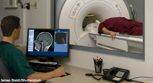
The U.S. Food and Drug Administration (FDA) is investigating the risk of brain deposits developing after repeated use of gadolinium-based contrast agents (GBCAs) for magnetic resonance imaging (MRI).1 Recent medical publications have reported that GBCA deposits have remained in patient brains long after the last administration of these agents, particularly in patients who have undergone at least four MRI scans. It is not known whether effects from the gadolinium deposits are harmful or if they can lead to adverse health effects.

FDA is continuing to further study any potential safety risks. No current changes to the product labeling for GBCAs are being recommended at this time.

ad goes here:advert-1
ADVERTISEMENT
SCROLL TO CONTINUE

To prevent the potential accumulation of gadolinium, the FDA is requesting that healthcare professionals limit GBCA use to clinical circumstances in which the additional information provided by the contrast is necessary. The FDA is also requesting that healthcare professionals reassess the necessity of repetitive GBCA MRIs in established treatment protocols.

In published studies, investigators reviewed noncontrast MRIs of patients who had received several GBCA MRIs as part of cancer management, multiple sclerosis management or for other illnesses. The noncontrast MRIs demonstrated findings that suggested gadolinium contrast was being retained in various brain structures. So far there have been no signs or symptoms of adverse health effects and no pathological changes associated with these gadolinium brain deposits. In some of these studies, brain examination at autopsy confirmed gadolinium deposits.

ad goes here:advert-2
ADVERTISEMENT
SCROLL TO CONTINUE

GBCAs include gadofosveset (Ablavar), gadoterate meglumine (Dotarem), gadoxetate disodium (Eovist), gadobutrol (Gadavist), gadopentetate dimeglumine (Magnevist), gadobenate dimeglumine (MultiHance), gadodiamide (Omniscan), gadoversetamide (Optimark) and gadoteridol (ProHance).

Michele B. Kaufman, PharmD, CGP, RPh, is a freelance medical writer based in New York City and a pharmacist at New York Presbyterian Lower Manhattan Hospital. 

Reference

  1. U.S. Food and Drug Administration. Drug safety communication: FDA evaluating the risk of brain deposits with repeated used of gadolinium-based contrast agents for magnetic resonance imaging (MRI). 2015 July 27.

Share: 

Filed under:Drug Updates Tagged with:brainFDAMagnetic resonance imaging (MRI)

Related Articles

    Rheumatology Drug Updates on Brentuximab Vedotin, Tofacitinib Citrate

    November 17, 2015

    Brentuximab Vedotin Enters Phase 2 Trials Brentuximab vedotin (Adcetris), an antibody-drug conjugate (ADC) directed at CD30, is currently entering Phase 2 clinical trials for treating systemic lupus erythematosus (SLE).1 The ADC encompasses an anti-CD30 monoclonal antibody, which is attached by a protease-cleavable linker to a microtubule-disrupting agent, known as monomethyl auristatin E (MMAE). The ADC…

    What Causes Nephrogenic Systemic Fibrosis?

    September 1, 2007

    Imaging studies and kidney problems may trigger this gadolinium-induced fibrosing syndrome

    Envision Arthritis Pathology

    April 1, 2008

    MRI advances in RA and OA

    Imaging in Ankylosing Spondylitis

    April 1, 2015

    MRI inflammation, fat and new bone formation in the sacroiliac joints, spine in patients with AS

  • About Us
  • Meet the Editors
  • Issue Archives
  • Contribute
  • Advertise
  • Contact Us
  • Copyright © 2025 by John Wiley & Sons, Inc. All rights reserved, including rights for text and data mining and training of artificial technologies or similar technologies. ISSN 1931-3268 (print). ISSN 1931-3209 (online).
  • DEI Statement
  • Privacy Policy
  • Terms of Use
  • Cookie Preferences