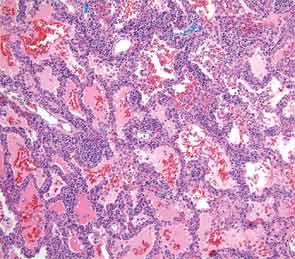
Video: Knock on Wood| Webinar: ACR/CHEST ILD Guidelines in Practice
fa-facebookfa-linkedinfa-youtube-playfa-rss

An official publication of the ACR and the ARP serving rheumatologists and rheumatology professionals

  • Conditions
    • Axial Spondyloarthritis
    • Gout and Crystalline Arthritis
    • Myositis
    • Osteoarthritis and Bone Disorders
    • Pain Syndromes
    • Pediatric Conditions
    • Psoriatic Arthritis
    • Rheumatoid Arthritis
    • Sjögren’s Disease
    • Systemic Lupus Erythematosus
    • Systemic Sclerosis
    • Vasculitis
    • Other Rheumatic Conditions
  • FocusRheum
    • ANCA-Associated Vasculitis
    • Axial Spondyloarthritis
    • Gout
    • Lupus Nephritis
    • Psoriatic Arthritis
    • Rheumatoid Arthritis
    • Systemic Lupus Erythematosus
  • Guidance
    • Clinical Criteria/Guidelines
    • Ethics
    • Legal Updates
    • Legislation & Advocacy
    • Meeting Reports
      • ACR Convergence
      • Other ACR meetings
      • EULAR/Other
    • Research Rheum
  • Drug Updates
    • Analgesics
    • Biologics/DMARDs
  • Practice Support
    • Billing/Coding
    • EMRs
    • Facility
    • Insurance
    • QA/QI
    • Technology
    • Workforce
  • Opinion
    • Patient Perspective
    • Profiles
    • Rheuminations
      • Video
    • Speak Out Rheum
  • Career
    • ACR ExamRheum
    • Awards
    • Career Development
  • ACR
    • ACR Home
    • ACR Convergence
    • ACR Guidelines
    • Journals
      • ACR Open Rheumatology
      • Arthritis & Rheumatology
      • Arthritis Care & Research
    • From the College
    • Events/CME
    • President’s Perspective
  • Search

Fellow’s Forum Case Report: Diagnosing Antisynthetase Syndrome

Fawad Aslam, MBBS, and Elizabeth B. Russell, MD  |  Issue: May 2013  |  May 1, 2013

An extensive infectious disease workup for viral infections, tuberculosis, fungi, and bacteria was negative. A transbronchial biopsy was negative for any infectious etiology or granulomas. Histopathology revealed nonspecific interstitial pneumonitis (NSIP) pattern. The otolaryngology service was consulted for a minor salivary gland biopsy, which was subsequently reported as being consistent with Sjögren’s syndrome (SS) based on more than one focus of more than 50 lymphocytes per 4 mm2 (see Figure 3). A surgical lung biopsy confirmed NSIP (see Figure 4); fibrosis was absent. At this time, the anti-Jo-1 antibody was reported as positive at 7.9 (0–0.9 AI)). The serum aldolase level was elevated at 9.4 (1.5–8.1 U/L).

A diagnosis of antisynthetase syndrome with subclinical SS—based on positive serologies and biopsy in the absence of sicca symptoms—was made. The patient was placed on oral prednisone at 1 mg/kg and hydroxychloroquine 200 milligrams twice daily. After three days of treatment, he had a remarkable response and his joint symptoms resolved. At three months of follow-up, he was on prednisone 40 mg, had no complaints, and his PFT and chest radiograph had normalized. He was started on methotrexate 10 mg weekly to allow for steroid taper.

ad goes here:advert-1
ADVERTISEMENT
SCROLL TO CONTINUE

Chest radiograph showing nodular opacities in both lungs.
Figure 1: Chest radiograph showing nodular opacities in both lungs.

ad goes here:advert-2
ADVERTISEMENT
SCROLL TO CONTINUE
CT scan of the lung showing multifocal areas of ground-glass and reticular opacities.
Figure 2: CT scan of the lung showing multifocal areas of ground-glass and reticular opacities.

Figure 3: Minor salivary glands with a dense lymphoplasmacytic infiltrate.

Marked interstitial chronic inflammatory infiltrate as well as scattered large lymphoid aggregates. Increased numbers of histiocytes are present within the alveolar spaces along with proteinaceous material. Biopsy findings are suggestive of NSIP.
Figure 4: Marked interstitial chronic inflammatory infiltrate as well as scattered large lymphoid aggregates. Increased numbers of histiocytes are present within the alveolar spaces along with proteinaceous material. Biopsy findings are suggestive of NSIP.

Discussion

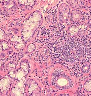
Rheumatologists are often asked to evaluate patients presenting with interstitial lung disease (ILD) in association with a positive ANA or RF, joint, or musculoskeletal symptoms. This case highlights one such typical scenario. The initial concern was for an ANCA-associated disease based on the presence of aural symptoms, hemoptysis, lung disease, and arthritis. However lack of ANCA antibody positivity, absence of any bony erosion on sinus imaging, and subsequent biopsy without histological evidence of vasculitis merited a diagnostic reevaluation.

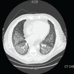
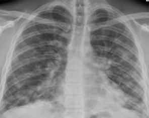
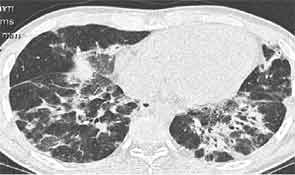
Antisynthetase syndrome is a systemic autoimmune syndrome characterized by a variable combination of ILD, myopathy, fever, joint involvement, and mechanic’s hands with a reported prevalence of 1.5 per 100,000. Its hallmark is the presence of antibodies directed against aminoacyl-transfer RNA synthetase of which the most frequent is the anti-Jo-1 antibody that accounts for about 80% of all cases of antisynthetase syndrome.1 Other relatively common antibodies include anti-PL-7 and anti-EJ.2 Anti-Jo-1 antibodies are present in about 30% of patients presenting with myositis and it is in the setting of myositis that these antibodies are usually tested to diagnose antisynthetase syndrome. ILD, however, is the most frequently occurring manifestation of antisynthetase syndrome and is reported in 75% to 89% of all cases.3 Lung involvement is the most important determinant of prognosis.

Page: 1 2 3 4 5 6 | Single Page
Share: 

Filed under:ConditionsOther Rheumatic Conditions Tagged with:Antisynthetase SyndromeInterstitial Lung Diseasepatient care

Related Articles

    What Rheumatologists Need to Know about Diagnosing and Managing Interstitial Lung Disease (ILD)

    December 1, 2012

    Patients with systemic sclerosis (SSc), poly-/dermatomyositis (PM/DM), or rheumatoid arthritis (RA) appear to carry the greatest risk for developing connective tissue disease-associated interstitial lung disease (CTD-ILD)
     

    How to Diagnose Antisynthetase Syndrome

    How to Diagnose Antisynthetase Syndrome

    March 17, 2017

    Antisynthetase syndrome (AS) is strongly associated with the presence of antibodies to aminoacyl-transfer RNA (tRNA) synthetases (ARSs) that are implicated in the pathogenesis of myositis and interstitial lung disease (ILD). Antibodies against eight antisynthetases have been identified and are detected in 16–26% of patients with idiopathic inflammatory myopathies (IIM).1 Serum assays for five of these…

    Scleroderma & ILD: Practical Tips on the Diagnosis & Management of Systemic Sclerosis-Associated Interstitial Lung Disease

    June 15, 2022

    No one-size-fits-all approach exists for the care and treatment of patients with systemic sclerosis (SSc) and SSc with pulmonary involvement. Here, experts discuss some best clinical practices for these patients.

    Fellow’s Forum Case Report: When Pulmonary Symptoms Point to Rheumatic Disease

    September 1, 2013

    A 48-year-old man with diffuse arthralgias and acute respiratory failure is diagnosed with antisynthetase syndrome

  • About Us
  • Meet the Editors
  • Issue Archives
  • Contribute
  • Advertise
  • Contact Us
fa-facebookfa-linkedinfa-youtube-playfa-rss
  • Copyright © 2025 by John Wiley & Sons, Inc. All rights reserved, including rights for text and data mining and training of artificial technologies or similar technologies. ISSN 1931-3268 (print). ISSN 1931-3209 (online).
  • DEI Statement
  • Privacy Policy
  • Terms of Use
  • Cookie Preferences