Video: Every Case Tells a Story| Webinar: ACR/CHEST ILD Guidelines in Practice

An official publication of the ACR and the ARP serving rheumatologists and rheumatology professionals

  • Conditions
    • Axial Spondyloarthritis
    • Gout and Crystalline Arthritis
    • Myositis
    • Osteoarthritis and Bone Disorders
    • Pain Syndromes
    • Pediatric Conditions
    • Psoriatic Arthritis
    • Rheumatoid Arthritis
    • Sjögren’s Disease
    • Systemic Lupus Erythematosus
    • Systemic Sclerosis
    • Vasculitis
    • Other Rheumatic Conditions
  • FocusRheum
    • ANCA-Associated Vasculitis
    • Axial Spondyloarthritis
    • Gout
    • Psoriatic Arthritis
    • Rheumatoid Arthritis
    • Systemic Lupus Erythematosus
  • Guidance
    • Clinical Criteria/Guidelines
    • Ethics
    • Legal Updates
    • Legislation & Advocacy
    • Meeting Reports
      • ACR Convergence
      • Other ACR meetings
      • EULAR/Other
    • Research Rheum
  • Drug Updates
    • Analgesics
    • Biologics/DMARDs
  • Practice Support
    • Billing/Coding
    • EMRs
    • Facility
    • Insurance
    • QA/QI
    • Technology
    • Workforce
  • Opinion
    • Patient Perspective
    • Profiles
    • Rheuminations
      • Video
    • Speak Out Rheum
  • Career
    • ACR ExamRheum
    • Awards
    • Career Development
  • ACR
    • ACR Home
    • ACR Convergence
    • ACR Guidelines
    • Journals
      • ACR Open Rheumatology
      • Arthritis & Rheumatology
      • Arthritis Care & Research
    • From the College
    • Events/CME
    • President’s Perspective
  • Search

Fellow’s Forum Case Report: When Pulmonary Symptoms Point to Rheumatic Disease

Naina Rastalsky, MD  |  Issue: September 2013  |  September 1, 2013

Given the presence of thickened cracked skin on the patient’s hands, which we felt to be consistent with mechanic’s hands, negative ANA, positive cytoplasmic and Ro antibodies, as well as history of Raynaud’s phenomenon and arthralgias, an antisynthetase syndrome was considered. A myositis-specific antibody panel was sent.

The patient then underwent a right upper-lobe wedge biopsy. This showed acute and organizing diffuse alveolar damage (DAD) with marked cytologic atypia, vascular thrombosis, collections of eosinophils, and vascular mural thickening. Hyaline membranes involved most of the specimen (see Figure 3). Immunohistochemistry for HSV showed a rare, weakly positive cell but no characteristic viral cytopathic inclusions. Immunohistochemistry for cytomegalovirus was negative. No organisms were seen on Grocott’s methenamine silver stain (GMS), AFB, Brown-Hopps, or Steiner stains. There was no evidence of malignancy.

ad goes here:advert-1
ADVERTISEMENT
SCROLL TO CONTINUE

Diffuse alveolar damage. Transition between less-involved lung and severely involved lung
Figure 3A: Diffuse alveolar damage. Transition between less-involved lung and severely involved lung.

ad goes here:advert-2
ADVERTISEMENT
SCROLL TO CONTINUE
Vascular mural thickening and organizing fibrosis
Figure 3B: Vascular mural thickening and organizing fibrosis.

Hyaline membranes define the pathologic finding of diffuse alveolar damage. Both acute (bright pink, thin membranes) and organizing (more cellular regions) phases are seen
Figure 3C: Hyaline membranes define the pathologic finding of diffuse alveolar damage. Both acute (bright pink, thin membranes) and organizing (more cellular regions) phases are seen.

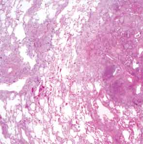
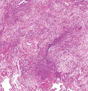
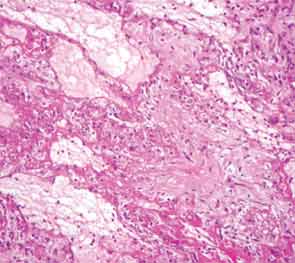
The overall picture was that of an acute alveolar injury in an exudative and fibroproliferative phase, superimposed on an older process with organizing pneumonia by perhaps two to three weeks. Some lymphoid aggregates were suggestive of possible autoimmune etiology.

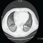
His lung biopsy was complicated by pneumothorax (see Figure 4). He required multiple chest tube placements. He underwent a tracheostomy procedure.

In mid January, two days after his lung biopsy, pulsed dose steroids were started, 1 g solumedrol IV daily for three days, followed by prednisone 60 mg daily. He was continued on acyclovir for prophylaxis for HSV reactivation. Because of persistent fevers, broad-spectrum antibiotics were resumed. Cyclophosphamide was considered, however, at the time, stains for pathogens from his lung biopsy were still pending. In this setting we worried about the added risk that a broad immunosuppressive approach with cyclophosphamide was going to pose and we chose instead to treat the patient with B-cell depletion only. He was thus started on rituximab and received 1 g IV on Day 2 of his pulsed steroid dose. A second dose of 1 g IV was given two weeks later. His respiratory status began to improve three days after the start of the immunosuppressive treatment. Over the ensuing week, he went on to have a dramatic clinical response and was able to wean off of ventilator support completely eight days after initiation of immunosuppressive treatment. The myositis-specific antibody panel returned positive for PL-7.

Page: 1 2 3 4 5 | Single Page
Share: 

Filed under:ConditionsSystemic Sclerosis Tagged with:Antisynthetase SyndromearthralgiasPulmonaryrespiratoryRheumatic Disease

Related Articles

    New Tools for Myositis Diagnosis, Classification & Management

    April 15, 2019

    CHICAGO—At Hot Topics in Myositis, a session at the 2018 ACR/ARHP Annual Meeting, three experts discussed new classification criteria for idiopathic inflammatory myopathies (IIM) and offered practical primers on overlap myositis conditions and inclusion body myositis (IBM). New Myositis Classification Criteria After a 10-year development process, the new EULAR/ACR Classification Criteria for Adult and Juvenile…

    Fellow’s Forum Case Report: Diagnosing Antisynthetase Syndrome

    May 1, 2013

    A variable combination of interstitial lung disease, myopathy, fever, joint involvement, and mechanic’s hands characterize this systemic autoimmune syndrome

    Autoantibodies in Autoimmune Myopathy

    Autoantibodies in Autoimmune Myopathy

    September 18, 2017

    In recent years, scientists and clinicians have learned a great deal about autoantibodies occurring in idiopathic inflammatory myopathies (IIMs). These new discoveries have reshaped our understanding of distinct clinical pheno­types in IIMs. Scientists continue to learn more about how these auto­antibodies shape pathophysiology, diagnosis, disease monitoring, prognosis and optimum treatment. Moving forward, these autoantibodies will…

    How to Diagnose Antisynthetase Syndrome

    How to Diagnose Antisynthetase Syndrome

    March 17, 2017

    Antisynthetase syndrome (AS) is strongly associated with the presence of antibodies to aminoacyl-transfer RNA (tRNA) synthetases (ARSs) that are implicated in the pathogenesis of myositis and interstitial lung disease (ILD). Antibodies against eight antisynthetases have been identified and are detected in 16–26% of patients with idiopathic inflammatory myopathies (IIM).1 Serum assays for five of these…

  • About Us
  • Meet the Editors
  • Issue Archives
  • Contribute
  • Advertise
  • Contact Us
  • Copyright © 2025 by John Wiley & Sons, Inc. All rights reserved, including rights for text and data mining and training of artificial technologies or similar technologies. ISSN 1931-3268 (print). ISSN 1931-3209 (online).
  • DEI Statement
  • Privacy Policy
  • Terms of Use
  • Cookie Preferences