New Africa / shutterstock.com
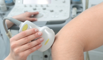
ATLANTA—Two ways to investigate injuries to the upper extremities are by in-depth physical examinations and ultrasound. In a Clinical Practice session at the 2019 ACR/ARP Annual Meeting, Anatomy: Correlating Physical Exam and Ultrasound in Common Sports Injuries of the Upper Extremity, Carlin Senter, MD, FACP, associate professor of primary care sports medicine at the University of California, San Francisco, and Anthony M. Reginato, MD, PhD, associate professor and director of the Division of Rheumatology at Brown University, Providence, R.I., presented strategies to diagnose different injuries via case studies.
Most sports medicine problems can be identified by conducting a careful history and physical exam and knowing your anatomy, said Dr. Senter. “However, musculoskeletal ultrasound is playing an increasingly important role in both diagnosis and guiding treatment for sports medicine problems.”
‘Musculoskeletal ultrasound is playing an increasingly important role in both diagnosis & guiding treatment for sports medicine problems.’ —Carlin Senter, MD, FACP
“In rheumatology, ultrasound can predict diagnosis—as well as provide early detection—staging of the disease and monitor response to therapy,” said Dr. Reginato.
A Case of Shoulder Pain
In one case, a 57-year-old man presented with right shoulder pain that started after he had slipped and fallen three months earlier. He tried physical therapy without benefit and was waking up at night due to pain. On physical exam, the shoulder was not tender. The active range of motion of the shoulder was intact, with pain on abduction between 60º and 120º. He had moderate pain with resisted external rotation of the shoulder and was unable to perform a lift-off test. The diagnosis was a rotator cuff tendon tear.

Dr. Senter
“Rotator cuff disease is the most common problem affecting adults with shoulder pain,” said Dr. Senter. “The painful arc test and testing the rotator cuff muscle strength using resisted shoulder external and internal rotation, as well as the empty can test, are useful in identifying patients with rotator cuff disease.”
Dr. Reginato said musculoskeletal ultrasound can identify all four types of rotator cuff disease—impingement, tendinopathy or tendinitis, partial-thickness tears and full-thickness tears.
Elbow Complaint
In another case, a 33-year-old right-handed man presented with three weeks of right lateral elbow pain. The pain came on gradually and worsened with grip activities. He had been remodeling his home and had trouble using a screwdriver and felt weak carrying groceries. He had no numbness or tingling in his arms or hands.
An exam showed that his neck was normal, and he did not have swelling or ecchymosis of elbow. There was tenderness at the lateral epicondyle and just distal/anterior to lateral epicondyle. The patient had full, symmetric, bilateral elbow range of motion and pain with resisted wrist extension. There was no laxity with varus or valgus stress to the elbow.
The diagnosis was lateral epicondylitis, the most common cause of lateral elbow pain.
The Most Common Nerve Entrapment
Another case concerned a 47-year-old woman with an eight-month history of numbness and tingling in both hands that mostly affected the palmar aspect of the thumbs, index and middle fingers. She worked as a postal worker, with repetitive hand twisting throughout the day that caused worsening numbness as the day went on.

Dr. Reginato
Examination of the small joints of the hand was normal, and the wrist exam showed no swelling or pain on palpation. She had a positive Tinel’s sign at the wrist and Phalen’s maneuver, and no intrinsic muscle wasting of the hands. She was diagnosed with carpal tunnel syndrome.
“This is the most common nerve entrapment,” said Dr. Senter. “There [are] data showing that ultrasound can aid in the diagnosis of carpal tunnel syndrome.”1-3
“With ultrasound,” said Dr. Reginato, “you investigate the flexor carpi radialis tendon, the palmaris longus tendon—which is absent in 12.7% of the population—the median nerve and the radial artery.” The transducer is aligned longitudinally to the fingers. “It’s important that you do a dynamic exploration; you’re not just looking at the entrance of the carpal tunnel.”
Wrist Pain
In another case, a 32-year-old right-handed woman presented with two weeks of right wrist pain. The pain was localized to the dorsoradial aspect of the wrist. She complained it was worse using the steering wheel, and unscrewing bottles, which she was doing frequently since having her first child three months previously. She had not experienced trauma and did not have numbness or tingling in her hand.
Upon examination, her point of maximal tenderness was at the radial styloid, and she had a positive Finkelstein’s test. She was diagnosed with De Quervain’s tenosynovitis, inflammation of the tendons of the first dorsal compartment of the wrist. “It’s found on physical exam by point tenderness of the radial styloid and increased pain with passive ulnar deviation of the wrist,” said Dr. Senter.
“The key things we look for when evaluating the extensor tendons of the wrist with ultrasound is to look at the first compartment of the wrist, primarily at the abductor pollicis longus and extensor pollicis longus,” said Dr. Reginato. “You might see a marked thickening and hypoechoic appearance of the retinaculum in both a transverse and longitudinal view.”
Both Dr. Senter and Dr. Reginato agree: Thorough physical exams by a knowledgeable therapist and ultrasound have roles in the diagnosis and treatment recommendations for sports medicine problems related to the musculoskeletal system,
Mike Fillon is a healthcare writer living in the Atlanta area.
References
- Dejaco C, Stradner M, Zauner D, et al. Ultrasound for diagnosis of carpal tunnel syndrome: Comparison of different methods to determine median nerve volume and value of power Doppler sonography. Ann Rheum Dis. 2013 Dec;72(12):1934–1939.
- Emril DR, Zakaria I, Amrya M. Agreement between high-resolution ultrasound and electro-physiological examinations for diagnosis of carpal tunnel syndrome in the Indonesian population. Front Neurol. 2019 Aug 26;10:888.
- Li M, Jiang J, Zhou Q, Zhang C. Sonographic follow-up after endoscopic carpal tunnel release for severe carpal tunnel syndrome: A one-year neuroanatomical prospective observational study. BMC Musculoskelet Disord. 2019 Apr 9;20(1):157.