Video: Every Case Tells a Story| Webinar: ACR/CHEST ILD Guidelines in Practice

An official publication of the ACR and the ARP serving rheumatologists and rheumatology professionals

  • Conditions
    • Axial Spondyloarthritis
    • Gout and Crystalline Arthritis
    • Myositis
    • Osteoarthritis and Bone Disorders
    • Pain Syndromes
    • Pediatric Conditions
    • Psoriatic Arthritis
    • Rheumatoid Arthritis
    • Sjögren’s Disease
    • Systemic Lupus Erythematosus
    • Systemic Sclerosis
    • Vasculitis
    • Other Rheumatic Conditions
  • FocusRheum
    • ANCA-Associated Vasculitis
    • Axial Spondyloarthritis
    • Gout
    • Psoriatic Arthritis
    • Rheumatoid Arthritis
    • Systemic Lupus Erythematosus
  • Guidance
    • Clinical Criteria/Guidelines
    • Ethics
    • Legal Updates
    • Legislation & Advocacy
    • Meeting Reports
      • ACR Convergence
      • Other ACR meetings
      • EULAR/Other
    • Research Rheum
  • Drug Updates
    • Analgesics
    • Biologics/DMARDs
  • Practice Support
    • Billing/Coding
    • EMRs
    • Facility
    • Insurance
    • QA/QI
    • Technology
    • Workforce
  • Opinion
    • Patient Perspective
    • Profiles
    • Rheuminations
      • Video
    • Speak Out Rheum
  • Career
    • ACR ExamRheum
    • Awards
    • Career Development
  • ACR
    • ACR Home
    • ACR Convergence
    • ACR Guidelines
    • Journals
      • ACR Open Rheumatology
      • Arthritis & Rheumatology
      • Arthritis Care & Research
    • From the College
    • Events/CME
    • President’s Perspective
  • Search

Imaging Modalities in Gout: How to Use them in Clinical Practice

Samantha C. Shapiro, MD  |  Issue: September 2022  |  July 21, 2022

DECT

When it comes to dual-energy computed tomography, Dr. Pascart noted that “diagnostic accuracy is a bit better for DECT than ultrasound, with the exception of early disease.” In early disease (i.e., diagnosis of gout within the first two years of disease onset), ultrasound is more sensitive for new deposits as appreciated by the double contour sign.5 This is because DECT is limited by resolution. Significant MSU crystal aggregates need to be present to see them.

It’s also important to make sure that radiologists aren’t counting artifact while calculating MSU crystal burden on DECT images. “Nailbeds have the same signature as MSU crystals and shouldn’t be counted. Metal implants can also cause artifact,” Dr. Pascart explained.

ad goes here:advert-1
ADVERTISEMENT
SCROLL TO CONTINUE

“I like to use DECT for prognosis and follow-up,” Dr. Pascart said, “but I do admit I’m a bit biased since it’s so cool. We know there’s a relationship between the volume of crystals measured with DECT at baseline and flare risk over the next six months.6 With ultrasound, after six months of treatment, patients with a greater than 50% decrease in tophus size had less risk of flaring after those six months of treatment.7 [Given this information], you might argue for a lower serum urate target or prolonged flare prophylaxis if tophus burden as measured by DECT remains high.”

Dual-energy computed tomography & ultrasound are both more sensitive than plain radiographs & provide noninvasive characterization of monosodium urate crystals with specificity.

Ultrasound & DECT

What about using ultrasound and DECT in combination? Dr. Pascart et al. used prospectively collected data from an outpatient rheumatology clinic to examine the diagnostic accuracy of either modality alone or in combination, by anatomical site (i.e., feet and ankles, and knees).5 “The general conclusion,” Dr. Pascart explained, “was that there was no advantage to combining the two techniques. You gained some sensitivity but lost some specificity.”

In Sum

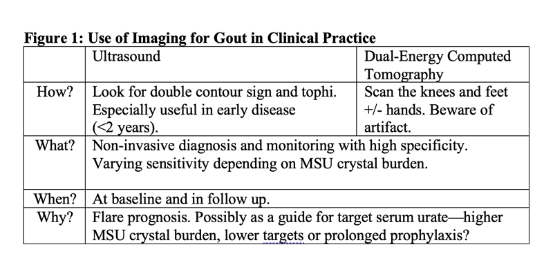
Dr. Pascart concluded his talk with a highyield summary of the “how, what, when and why” of imaging modalities in gout (see Figure 1, below). As ultrasound and DECT become more widely available, we can all hope for better care of our gout patients in the future.


Samantha C. Shapiro, MD, is an academic rheumatologist and an affiliate faculty member of the Dell Medical School at the University of Texas at Austin. She is also a member of the ACR Insurance Subcommittee.
ad goes here:advert-2
ADVERTISEMENT
SCROLL TO CONTINUE

Page: 1 2 3 | Single Page
Share: 

Filed under:ConditionsEULAR/OtherGout and Crystalline ArthritisMeeting Reports Tagged with:DECTdual-energy computed tomography (DECT)EULARGoutGout Resource Centerimagingmonosodium urate crystalsUltrasound

Related Articles

    Clinical Applications of Dual-Energy Computed Tomography for Rheumatology

    June 1, 2014

    Advanced imaging technique allows physicians to detect deposition of monosodium urate crystals not apparent in physical exams and better diagnose gout

    Difficult Gout

    July 1, 2007

    “Grandpapa’s Torments” was the Rodnan Commemorative Gout Print featured at the 2005 ACR/ARHP Annual Scientific Meeting.

    Gout, Glucose Metabolism and Obesity: A Case Review

    November 2, 2014

    New research explores association between hyperurecimia and gout with metabolic derangement

    Metabolic Biosensor Implicated in Gout

    February 1, 2015

    AMP-activated protein kinase may be link between nutritional excess, inflammation that leads to gout

  • About Us
  • Meet the Editors
  • Issue Archives
  • Contribute
  • Advertise
  • Contact Us
  • Copyright © 2025 by John Wiley & Sons, Inc. All rights reserved, including rights for text and data mining and training of artificial technologies or similar technologies. ISSN 1931-3268 (print). ISSN 1931-3209 (online).
  • DEI Statement
  • Privacy Policy
  • Terms of Use
  • Cookie Preferences