Video: Every Case Tells a Story| Webinar: ACR/CHEST ILD Guidelines in Practice

An official publication of the ACR and the ARP serving rheumatologists and rheumatology professionals

  • Conditions
    • Axial Spondyloarthritis
    • Gout and Crystalline Arthritis
    • Myositis
    • Osteoarthritis and Bone Disorders
    • Pain Syndromes
    • Pediatric Conditions
    • Psoriatic Arthritis
    • Rheumatoid Arthritis
    • Sjögren’s Disease
    • Systemic Lupus Erythematosus
    • Systemic Sclerosis
    • Vasculitis
    • Other Rheumatic Conditions
  • FocusRheum
    • ANCA-Associated Vasculitis
    • Axial Spondyloarthritis
    • Gout
    • Psoriatic Arthritis
    • Rheumatoid Arthritis
    • Systemic Lupus Erythematosus
  • Guidance
    • Clinical Criteria/Guidelines
    • Ethics
    • Legal Updates
    • Legislation & Advocacy
    • Meeting Reports
      • ACR Convergence
      • Other ACR meetings
      • EULAR/Other
    • Research Rheum
  • Drug Updates
    • Analgesics
    • Biologics/DMARDs
  • Practice Support
    • Billing/Coding
    • EMRs
    • Facility
    • Insurance
    • QA/QI
    • Technology
    • Workforce
  • Opinion
    • Patient Perspective
    • Profiles
    • Rheuminations
      • Video
    • Speak Out Rheum
  • Career
    • ACR ExamRheum
    • Awards
    • Career Development
  • ACR
    • ACR Home
    • ACR Convergence
    • ACR Guidelines
    • Journals
      • ACR Open Rheumatology
      • Arthritis & Rheumatology
      • Arthritis Care & Research
    • From the College
    • Events/CME
    • President’s Perspective
  • Search

How Magnetic Resonance Imaging Technology Can Aid Spondyloarthritis Diagnosis

Larry Beresford  |  Issue: August 2016  |  August 11, 2016

“We combine and synthesize information from the T1 and STIR scans to make a contextual interpretation of the information provided by MRI. Both sequences are important to diagnosis,” he said. The unfolding of events in the sacroiliac joint typically starts with inflammation just beneath the subchondral bone. After the inflammation has resolved, then what’s left is erosion of bone and fat metaplasia, a feature of repair in spondyloarthritis. “Then we see the development of new bone.” There is a difference between physiological fat and pathological fat metaplasia, which is distinguished on the MRI from normal, physiological fat by the presence of a distinct border, homogeneity, and proximity to subchondral bone, Dr. Maksymowych explained. This fat metaplasia is a key intermediary in the pathway from inflammation to the development of sacroiliac joint ankylosis on the MRI.

“The T1 sequence gives excellent detail around anatomy—beautiful contrast between cortical bone and underlying marrow,” he said, while the STIR allows identification of inflammation. Together, the two image sequences give information about where the inflammation is, the presence of repair tissue and erosion of bone. “All of these things together should enhance your confidence in the diagnosis.”

ad goes here:advert-1
ADVERTISEMENT
SCROLL TO CONTINUE

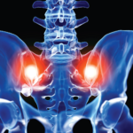
This kind of MRI-facilitated diagnosis can also have an impact on the patient, Dr. Maksymowych said—especially if the rheumatologist is able to show the visual evidence of active disease on the MRI images during the office visit. “It is an effective way of making the point that people need to take their medications if they want effective control of this disease. When they see the actual picture, it can have a huge impact on the problem of adherence to medication,” he explained.

“But the effective use of MRI in routine clinical practice for evaluation of spondyloarthritis requires a specific imaging protocol and hands-on training for both rheumatologists and radiologists,” Dr. Maksymowych said. “Basic aspects of MRI should be a mandatory component of the rheumatology curriculum.”

ad goes here:advert-2
ADVERTISEMENT
SCROLL TO CONTINUE

Larry Beresford is an Oakland, Calif.-based freelance medical journalist.

Page: 1 2 3 | Single Page
Share: 

Filed under:Axial SpondyloarthritisConditionsResearch RheumTechnology Tagged with:California Rheumatology Alliance 2016ClinicalDiagnosismagnetic resonance imagingMRIoutcomepatient careResearchTechnology

Related Articles

    Imaging in Ankylosing Spondylitis

    April 1, 2015

    MRI inflammation, fat and new bone formation in the sacroiliac joints, spine in patients with AS

    Imaging of Axial Psoriatic Arthritis

    May 9, 2022

    The axial phenotype of psoriatic arthritis (axPsA) is an excellent example of a major controversy in rheumatology that has become the focus of attention because of the emergence of new therapies with different mechanisms of action for alleviating joint inflammation. It was first described in 1961 but, until recently, it has largely remained under the…

    Rheumatologists Make Progress Defining Spectrum of Axial Spondyloarthritis

    May 1, 2014

    New research clarifies terminology used for diagnosis, but questions remain around epidemiology, genetics and management of patients with axial skeletal inflammation

    Envision Arthritis Pathology

    April 1, 2008

    MRI advances in RA and OA

  • About Us
  • Meet the Editors
  • Issue Archives
  • Contribute
  • Advertise
  • Contact Us
  • Copyright © 2025 by John Wiley & Sons, Inc. All rights reserved, including rights for text and data mining and training of artificial technologies or similar technologies. ISSN 1931-3268 (print). ISSN 1931-3209 (online).
  • DEI Statement
  • Privacy Policy
  • Terms of Use
  • Cookie Preferences