Video: Every Case Tells a Story| Webinar: ACR/CHEST ILD Guidelines in Practice

An official publication of the ACR and the ARP serving rheumatologists and rheumatology professionals

  • Conditions
    • Axial Spondyloarthritis
    • Gout and Crystalline Arthritis
    • Myositis
    • Osteoarthritis and Bone Disorders
    • Pain Syndromes
    • Pediatric Conditions
    • Psoriatic Arthritis
    • Rheumatoid Arthritis
    • Sjögren’s Disease
    • Systemic Lupus Erythematosus
    • Systemic Sclerosis
    • Vasculitis
    • Other Rheumatic Conditions
  • FocusRheum
    • ANCA-Associated Vasculitis
    • Axial Spondyloarthritis
    • Gout
    • Psoriatic Arthritis
    • Rheumatoid Arthritis
    • Systemic Lupus Erythematosus
  • Guidance
    • Clinical Criteria/Guidelines
    • Ethics
    • Legal Updates
    • Legislation & Advocacy
    • Meeting Reports
      • ACR Convergence
      • Other ACR meetings
      • EULAR/Other
    • Research Rheum
  • Drug Updates
    • Analgesics
    • Biologics/DMARDs
  • Practice Support
    • Billing/Coding
    • EMRs
    • Facility
    • Insurance
    • QA/QI
    • Technology
    • Workforce
  • Opinion
    • Patient Perspective
    • Profiles
    • Rheuminations
      • Video
    • Speak Out Rheum
  • Career
    • ACR ExamRheum
    • Awards
    • Career Development
  • ACR
    • ACR Home
    • ACR Convergence
    • ACR Guidelines
    • Journals
      • ACR Open Rheumatology
      • Arthritis & Rheumatology
      • Arthritis Care & Research
    • From the College
    • Events/CME
    • President’s Perspective
  • Search

The Classification & Diagnosis of Granulomatosis with Polyangiitis

Harry E. Subramanian, Ravi Sutaria, MD, & Fotios Koumpouras, MD  |  Issue: August 2018  |  August 16, 2018

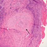
A repeat TEE demonstrated a vegetation on the mitral valve and thickened mitral valve leaflets, with mild regurgitation at the aortic, mitral and tricuspid valves. A skin biopsy of a periungual lesion demonstrated epidermal necrosis with acute inflammation and thrombi present, but vasculitis was not identified.

A magnetic resonance imaging (MRI) scan of the brain did not reveal any evidence of central nervous system vasculitis. A renal biopsy demonstrated necrotizing glomerulonephritis with crescents and without immune complex deposits, which was consistent with an ANCA-mediated glomerulonephritis (see Figure 2). A repeat bronchoscopy with bronchoalveolar lavage revealed active bleeding from the right upper lobe that was concerning for diffuse alveolar hemorrhage.

ad goes here:advert-1
ADVERTISEMENT
SCROLL TO CONTINUE

We determined the patient’s presentation was most consistent with GPA, and treatment was initiated with 375 mg/m2 of rituximab once per week for four weeks. In addition, plasma exchange was resumed, of which three cycles were completed (a total of five cycles).

Despite receiving extensive immunosuppressive treatments, the patient’s renal function continued to worsen, necessitating placement on regular hemodialysis. Further blood cultures and a lower respiratory culture did not grow any organism. Infectious disease tests for Bartonella henselae IgG and IgM, Coxiella burnetii IgG phases I and II, Brucella antibodies, HIV antibodies and DNA, hepatitis C virus antibodies and quantiferon-gold tuberculosis (TB) were negative.

ad goes here:advert-2
ADVERTISEMENT
SCROLL TO CONTINUE

Twenty-nine days after her initial presentation, the patient was extubated, but maintained a persistent oxygen requirement. The patient’s steroid dose was slowly tapered over her hospital course, eventually switching from methylprednisolone to prednisone, and down to a prednisone dose of 40 mg/day. During her recovery after extubation, it became evident that she was severely deconditioned and unable to leave her bed. She also had difficulty swallowing and suffered from multiple aspiration events, requiring placement of a gastrostomy tube for feeding.

Forty-nine days after her initial presentation, the patient developed respiratory distress thought to be secondary to aspiration, but declined any escalation in care, and she expired shortly thereafter. The patient’s family declined an autopsy.

Discussion

We present the case of a patient with GPA, whose clinical and laboratory findings were consistent with both an infective endocarditis and a small vessel vasculitis. The positive C-ANCA and PR3 antibody found during this patient’s workup did not provide any utility in discerning between these two diseases. The similarity in presentation between patients with ANCA-positive infective endocarditis and ANCA-associated vasculitis with endocardial involvement has been documented in the literature, and these cases are most often associated with a positive C-ANCA or PR3 antibody.7

Page: 1 2 3 4 5 6 7 8 | Single Page
Share: 

Filed under:ConditionsVasculitis Tagged with:ANCAantineutrophil cytoplasmic antibodyGPAgranulomatosis with polyangiitis

Related Articles
    Figure 1. A nasal biopsy shows intimal infiltration of the small blood vessels (black arrow).

    Case Illustrates the Difficulty Diagnosing Granulomatosis with Polyangiitis

    June 21, 2018

    Granulomatosis with polyangiitis (GPA) was first described in the British Medical Journal in 1897 by Scottish otolaryngologist Peter McBride.1 GPA is a relatively rare, systemic necrotizing vasculitis that can make diagnosis challenging. The incidence has been estimated anywhere between two and 12 cases per million.2 GPA mainly affects adults between the ages of 45 and…

    Case Report: A Patient on Apremilast Develops Streptococcus Salivarius

    October 18, 2019

    Apremilast was first marketed in March 2014 for the treatment of adults with psoriatic arthritis (PsA). An immuno­modulating drug, which is a small molecule inhibitor of phosphodiesterase 4 (PDE4) specific for cyclic adenosine mono­phosphate (cAMP), apremilast is administered orally. By inhibiting PDE4, intracellular cAMP levels are increased. Although the exact mechanism of action is not…

    Rheumatology’s Architect

    March 1, 2008

    Help the REF lay foundations for our future

    The Granulomatosis of Wegener’s

    May 16, 2011

    Delving deeper into the nonvasculitis aspects of the disease

  • About Us
  • Meet the Editors
  • Issue Archives
  • Contribute
  • Advertise
  • Contact Us
  • Copyright © 2025 by John Wiley & Sons, Inc. All rights reserved, including rights for text and data mining and training of artificial technologies or similar technologies. ISSN 1931-3268 (print). ISSN 1931-3209 (online).
  • DEI Statement
  • Privacy Policy
  • Terms of Use
  • Cookie Preferences