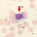
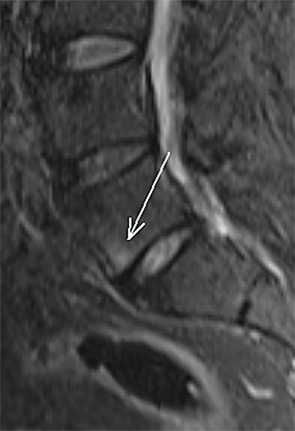
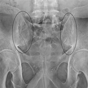
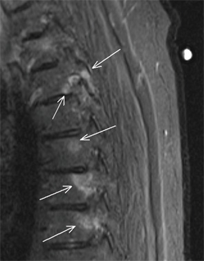
Video: Every Case Tells a Story| Webinar: ACR/CHEST ILD Guidelines in Practice

An official publication of the ACR and the ARP serving rheumatologists and rheumatology professionals

  • Conditions
    • Axial Spondyloarthritis
    • Gout and Crystalline Arthritis
    • Myositis
    • Osteoarthritis and Bone Disorders
    • Pain Syndromes
    • Pediatric Conditions
    • Psoriatic Arthritis
    • Rheumatoid Arthritis
    • Sjögren’s Disease
    • Systemic Lupus Erythematosus
    • Systemic Sclerosis
    • Vasculitis
    • Other Rheumatic Conditions
  • FocusRheum
    • ANCA-Associated Vasculitis
    • Axial Spondyloarthritis
    • Gout
    • Psoriatic Arthritis
    • Rheumatoid Arthritis
    • Systemic Lupus Erythematosus
  • Guidance
    • Clinical Criteria/Guidelines
    • Ethics
    • Legal Updates
    • Legislation & Advocacy
    • Meeting Reports
      • ACR Convergence
      • Other ACR meetings
      • EULAR/Other
    • Research Rheum
  • Drug Updates
    • Analgesics
    • Biologics/DMARDs
  • Practice Support
    • Billing/Coding
    • EMRs
    • Facility
    • Insurance
    • QA/QI
    • Technology
    • Workforce
  • Opinion
    • Patient Perspective
    • Profiles
    • Rheuminations
      • Video
    • Speak Out Rheum
  • Career
    • ACR ExamRheum
    • Awards
    • Career Development
  • ACR
    • ACR Home
    • ACR Convergence
    • ACR Guidelines
    • Journals
      • ACR Open Rheumatology
      • Arthritis & Rheumatology
      • Arthritis Care & Research
    • From the College
    • Events/CME
    • President’s Perspective
  • Search

The Diagnostic View: Our Findings

Jennifer L. Demertzis, MD  |  Issue: April 2015  |  April 1, 2015

Findings/Diagnosis

A posteroanterior supine radiograph of the sacroiliac (SI) joints (see Figure 1) demonstrates bilateral symmetric erosions of the SI joints without ankylosis and mild reactive sclerosis of both iliac bones adjacent to the joints (black circles). Review of thoracic and lumbar spine MRI examinations obtained two years prior to presentation show scattered areas of vertebral body bone marrow edema, centered at the vertebral body endplates and pedicles in the thoracic spine (see Figure 2, arrows) and in the anteroinferior corner of the L5 vertebral body in the lumbar spine (see Figure 3, arrow). Although initially interpreted as nonspecific, the MRI findings are consistent with spondylitis.

Based on the previous MRI findings of spondylitis and the current radiographic presence of symmetric erosive sacroiliitis, a diagnosis of ankylosing spondylitis was made. Subsequent HLA-B27 testing was positive, and the patient was started on therapy.

ad goes here:advert-1
ADVERTISEMENT
SCROLL TO CONTINUE

Figure 1

Figure 1

Figure 2

Figure 2


Figure 3

Figure 3

Take the challenge.


Jennifer L. Demertzis, MD, is a musculoskeletal radiologist at the Mallinckrodt Institute of Radiology at Washington University School of Medicine in St. Louis, Mo. She is excited to collaborate on this new feature in the journal and looks forward to seeing future cases contributed by readers.

ad goes here:advert-2
ADVERTISEMENT
SCROLL TO CONTINUE

Share: 

Filed under:Axial SpondyloarthritisConditions Tagged with:Ankylosing SpondylitisDemertzisdiagnosticdiagnostic imagingimagingPainradiographrheumatologyspine MRsymmetric erosive sacroiliitis

Related Articles

    Imaging in Ankylosing Spondylitis

    April 1, 2015

    MRI inflammation, fat and new bone formation in the sacroiliac joints, spine in patients with AS

    Case Report: Rheumatoid Arthritis Complicated by Large Granular Lymphocytic Leukemia

    November 14, 2021

    Figure 1. Peripheral blood smear showing a large granular lymphocyte in a 53-year-old Hispanic man. Large granular lymphocytic (LGL) leukemia is a rare, chronic, lymphoproliferative disorder of cytotoxic T cell or natural killer cell lineage with an annual incidence of 0.72 cases per 1 million people in the U.S.1 The most common sub­type of LGL…

    Meet the Lumbar Spinal Stenosis Challenge

    August 1, 2007

    Limited evidence and diagnostic options make this increasing condition difficult to treat

    Imaging of Axial Psoriatic Arthritis

    May 9, 2022

    The axial phenotype of psoriatic arthritis (axPsA) is an excellent example of a major controversy in rheumatology that has become the focus of attention because of the emergence of new therapies with different mechanisms of action for alleviating joint inflammation. It was first described in 1961 but, until recently, it has largely remained under the…

  • About Us
  • Meet the Editors
  • Issue Archives
  • Contribute
  • Advertise
  • Contact Us
  • Copyright © 2025 by John Wiley & Sons, Inc. All rights reserved, including rights for text and data mining and training of artificial technologies or similar technologies. ISSN 1931-3268 (print). ISSN 1931-3209 (online).
  • DEI Statement
  • Privacy Policy
  • Terms of Use
  • Cookie Preferences