Video: Every Case Tells a Story| Webinar: ACR/CHEST ILD Guidelines in Practice

An official publication of the ACR and the ARP serving rheumatologists and rheumatology professionals

  • Conditions
    • Axial Spondyloarthritis
    • Gout and Crystalline Arthritis
    • Myositis
    • Osteoarthritis and Bone Disorders
    • Pain Syndromes
    • Pediatric Conditions
    • Psoriatic Arthritis
    • Rheumatoid Arthritis
    • Sjögren’s Disease
    • Systemic Lupus Erythematosus
    • Systemic Sclerosis
    • Vasculitis
    • Other Rheumatic Conditions
  • FocusRheum
    • ANCA-Associated Vasculitis
    • Axial Spondyloarthritis
    • Gout
    • Psoriatic Arthritis
    • Rheumatoid Arthritis
    • Systemic Lupus Erythematosus
  • Guidance
    • Clinical Criteria/Guidelines
    • Ethics
    • Legal Updates
    • Legislation & Advocacy
    • Meeting Reports
      • ACR Convergence
      • Other ACR meetings
      • EULAR/Other
    • Research Rheum
  • Drug Updates
    • Analgesics
    • Biologics/DMARDs
  • Practice Support
    • Billing/Coding
    • EMRs
    • Facility
    • Insurance
    • QA/QI
    • Technology
    • Workforce
  • Opinion
    • Patient Perspective
    • Profiles
    • Rheuminations
      • Video
    • Speak Out Rheum
  • Career
    • ACR ExamRheum
    • Awards
    • Career Development
  • ACR
    • ACR Home
    • ACR Convergence
    • ACR Guidelines
    • Journals
      • ACR Open Rheumatology
      • Arthritis & Rheumatology
      • Arthritis Care & Research
    • From the College
    • Events/CME
    • President’s Perspective
  • Search

Using Ultrasound to Diagnose Carpal Tunnel Syndrome

Mark H. Greenberg, MD, RMSK, RhMSUS, Julian Greer, & James W. Fant Jr., MD  |  Issue: April 2018  |  April 26, 2018

Ultrasound evaluation may identify the etiology of median nerve impingement by recognizing structural signs, such as severe tenosynovitis, bifid median nerve, a persistent median artery, a ganglion cyst, a neoplasm, a lipoma, accessory muscles and calcium/uric acid/amyloid deposits. US may also demonstrate a vastly swollen median nerve, which increases the probability of injury from injection without US guidance.21

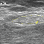
Tenosynovitis appears as a hypoechoic halo surrounding a tendon due to either thickening of the tenosynovium, fluid accumulation or both. Compression with the transducer to look for fluid displacement may help differentiate tenosynovitis from effusion.

ad goes here:advert-1
ADVERTISEMENT
SCROLL TO CONTINUE

In our clinic, we routinely perform an evaluation of the ulnar nerve in the ulnar canal because injury to the deep (motor) branch may cause hand weakness (including the first digit) and potentially mimic median neuropathy.

Carpal Tunnel Treatment?

Treatment of CTS includes risk factor mitigation, cock-up splints to prevent night-time wrist flexion, medication to treat inflammation, corticosteroids orally or by injection to mitigate inflammation, surgical decompression with transection of the TCL and removal of offending structures, such as cysts.

ad goes here:advert-2
ADVERTISEMENT
SCROLL TO CONTINUE

When injection is an option, US evaluation has been proved to increase the efficacy and safety of carpal tunnel injection and may help direct the optimal injection location.22 The normal anatomy of the carpal tunnel contents may be distorted due to disease process. Ultrasound can identify anatomic alteration, thereby preventing further damage to the median nerve. As seen with our patient, blind injection using usual topographic landmarks risked impaling the median nerve. Ultrasound was necessary to visualize significant FPL tenosynovitis that resulted in migration of the median nerve to a distinctly abnormal position, one that would subject it to damage with a topographic landmark-guided injection.

By delineating the etiology of the CTS, US may inform overall treatment decisions. There’s an opportunity for conservative treatment when US reveals the presence of significant tenosynovitis causing median nerve compression, as in our case. You can target tenosynovitis with a combination of medication directed at the underlying disease process (rheumatoid arthritis, in our patient’s case) as well as US-guided injections directly into the area or areas of flexor tenosynovitis that appear to be compressing the median nerve. We also recommended continued use of cock-up splints at night in our case.

US may also prove useful after surgery because surgery doesn’t always alleviate symptoms. Continued or recurrent symptoms may be due to iatrogenic nerve injury, incomplete TCL release, perineural fibrosis or severe presurgical damage to the median nerve.23 Ultrasound’s capacity to visualize these complications may prove useful in delineating the cause of persistent symptoms after surgery.

Conclusion

Ultrasound has become an important tool for CTS evaluation. This inexpensive and noninvasive technique offers criteria to confirm a clinical diagnosis, and it demonstrates morphology that helps avoid treatment pitfalls.

Page: 1 2 3 4 5 6 | Single Page
Share: 

Filed under:ConditionsSoft Tissue Pain Tagged with:Carpal Tunnel Syndromecase reportelectrophysiological studiesUltrasound

Related Articles
    Musculoskeletal Ultrasound: A Valuable Tool for Diagnosing Rheumatic Illnesses

    Musculoskeletal Ultrasound: A Valuable Tool for Diagnosing Rheumatic Illnesses

    October 15, 2015

    Musculoskeletal (MSK) ultrasound is a valuable imaging modality for the practicing rheumatologist and provides an efficient tool with high diagnostic value in the evaluation of patients with musculoskeletal complaints. The use of MSK ultrasound has evolved in the U.S. due to the emergence of less-expensive, portable ultrasound units, which provide high-quality gray-scale and power Doppler…

    Patients with Non-Classic Carpal Tunnel Syndrome Can Benefit from Surgery

    September 19, 2014

    Patients with extra-median spread of CTS symptoms had similar post-surgical outcomes as patients with classic CTS

    Patients with Parkinson’s Disease Are at Risk for Carpal Tunnel Syndrome

    November 21, 2016

    Patients with Parkinson’s disease may be at risk of carpal tunnel syndrome (CTS). New research examined why patients with Parkinson’s have a higher incidence of CTS than that of the general population. The study found that those patients who underwent subthalamic nucleus deep brain stimulation to monitor them for neuropathy may develop CTS…

    Right: The same view as 2A, with the common peroneal nerve outlined in yellow with a cross-sectional area of 21 mm2.

    Case Report: Ultrasound Reveals Cause of Post-Arthroplasty Knee Pain

    April 15, 2020

    A 65-year-old woman was referred by an orthopedist to a rheumatologist for left knee pain. Previously, in 2014, she underwent left total knee arthroplasty (TKA) for severe osteoarthritis in a different institution. Following the procedure, she experienced severe chronic anterolateral knee pain at rest, exacerbated by walking. Because she was rendered wheelchair bound and required…

  • About Us
  • Meet the Editors
  • Issue Archives
  • Contribute
  • Advertise
  • Contact Us
  • Copyright © 2025 by John Wiley & Sons, Inc. All rights reserved, including rights for text and data mining and training of artificial technologies or similar technologies. ISSN 1931-3268 (print). ISSN 1931-3209 (online).
  • DEI Statement
  • Privacy Policy
  • Terms of Use
  • Cookie Preferences