Video: Every Case Tells a Story| Webinar: ACR/CHEST ILD Guidelines in Practice

An official publication of the ACR and the ARP serving rheumatologists and rheumatology professionals

  • Conditions
    • Axial Spondyloarthritis
    • Gout and Crystalline Arthritis
    • Myositis
    • Osteoarthritis and Bone Disorders
    • Pain Syndromes
    • Pediatric Conditions
    • Psoriatic Arthritis
    • Rheumatoid Arthritis
    • Sjögren’s Disease
    • Systemic Lupus Erythematosus
    • Systemic Sclerosis
    • Vasculitis
    • Other Rheumatic Conditions
  • FocusRheum
    • ANCA-Associated Vasculitis
    • Axial Spondyloarthritis
    • Gout
    • Psoriatic Arthritis
    • Rheumatoid Arthritis
    • Systemic Lupus Erythematosus
  • Guidance
    • Clinical Criteria/Guidelines
    • Ethics
    • Legal Updates
    • Legislation & Advocacy
    • Meeting Reports
      • ACR Convergence
      • Other ACR meetings
      • EULAR/Other
    • Research Rheum
  • Drug Updates
    • Analgesics
    • Biologics/DMARDs
  • Practice Support
    • Billing/Coding
    • EMRs
    • Facility
    • Insurance
    • QA/QI
    • Technology
    • Workforce
  • Opinion
    • Patient Perspective
    • Profiles
    • Rheuminations
      • Video
    • Speak Out Rheum
  • Career
    • ACR ExamRheum
    • Awards
    • Career Development
  • ACR
    • ACR Home
    • ACR Convergence
    • ACR Guidelines
    • Journals
      • ACR Open Rheumatology
      • Arthritis & Rheumatology
      • Arthritis Care & Research
    • From the College
    • Events/CME
    • President’s Perspective
  • Search

Watch Those Eyes

Sirichai Pasadhika,MD and James T. Rosenbaum, MD  |  Issue: December 2007  |  December 1, 2007

Although ophthalmologists and rheumatologists come from the same medical-school background, our training can diverge so much that even the language we speak is incompletely shared. We know many rheumatologists who stare uncertainly at the notations that are the standard nomenclature for an eye examination. And we know ophthalmologists who are unfamiliar with the new drugs—including the biologics—that are now the mainstay of immunosuppressive therapy. Given the complexity of systemic inflammatory diseases, miscommunication about ocular problems can have serious consequences, causing delays in diagnoses and even misdirected therapy.

Case for Collaboration

Consider, for example, the management issues raised when the the Uveitis Clinic Casey Eye Institute at Oregon Health and Science University (OHSU) in Portland recently evaluated a patient with Wegener’s granulomatosis for retinal vasculitis. The patient was a 55-year-old female with biopsy-proven Wegener’s that had been treated with prednisone and cyclophosphamide. When the patient began to develop visual loss in one eye, her Wegener’s was seemingly in remission. The dilated ophthalmologic examination, however, demonstrated widespread retinal vasculitis. This examination also revealed large patches of creamy retinal injury that are characteristic of acute retinal necrosis (ARN), a herpetic infection that is rare but much more common in an immunocompromised host (see Figure 1).

ad goes here:advert-1
ADVERTISEMENT
SCROLL TO CONTINUE

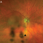
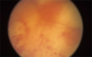
figure 1: Acute retinal necrosis from herpes virus infection.

The rheumatologic perspective was important in this case because we know that patients with a systemic vasculitis, such as polyarteritis or Wegener’s granulomatosis, rarely develop retinal vasculitis from the disease itself—especially if adequately treated. The ophthalmology perspective was equally important because the diagnosis of ARN is usually made by clinical examination that requires indirect ophthalmoscopy to identify the characteristic lesions. These lesions are often in the peripheral retina. Polymerase chain reaction (PCR) tests for a specific virus using surgically obtained ocular fluid can confirm the diagnosis. By amalgamating expertise in rheumatology and ophthalmology, we were able to appropriately diagnose this patient. The proper treatment of ARN is to reduce immunosuppression and initiate anti-viral therapy.

Ophthalmology Refresher

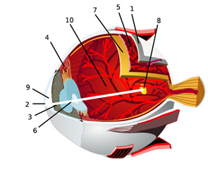
In an effort to facilitate communications with our ophthalmologic peers, this article will review the anatomy of the eye and the salient types of involvement in major rheumatologic diseases. (See Figure 2, for an overview of ocular anatomy.)

figure 2: Ocular Anatomy The eye consists of three layers. The outermost layer is the sclera (1) and cornea (2). While the sclera maintains the integrity of the globe, the clear cornea at the front of the eye refracts light into the eye.The middle layer is called the uvea, which consists of the iris (3), ciliary body (4), and choroids (5). Iris pigment determines eye color. The iris reacts to light like a camera shutter. The ciliary body secretes aqueous humor, and helps suspend the crystalline lens (6). The choroid lies in close proximity to the neural retina (7). The uvea is richly supplied with blood. An inflammation of the uvea is termed uveitis, but this term is also used to describe inflammation adjacent to the uvea. Vitritis, for example, is considered a subset of uveitis.s
ad goes here:advert-2
ADVERTISEMENT
SCROLL TO CONTINUE

Page: 1 2 3 4 5 6 7 8 9 | Single Page
Share: 

Filed under:Axial SpondyloarthritisBiologics/DMARDsConditionsOther Rheumatic Conditions Tagged with:BiologicsCorticosteroidsDiagnostic Criteriaimmunosuppressive drugsPathogenesisTreatmentUveitis

Related Articles

    An Overview of Pediatric, Noninfectious Uveitis

    October 18, 2018

    Uveitis is an inflammation of the uvea, which comprises the iris, ciliary body and choroid. Uveitis can lead to ocular damage and complete visual loss. Noninfectious etiologies for uveitis are the most common in the U.S.1 The estimated incidence of uveitis ranges from 25–52 per 100,000 in adults and five per 100,000 in children. The…

    ARZTSAMUI / shutterstock.com

    Uveitis: A Brief Primer for the Rheumatologist

    November 12, 2020

    Uveitis is an umbrella term for intraocular inflammatory diseases that can lead to vision loss. It’s not just a concern for ophthalmologists. Uveitis occurs in approximately 2–5% of patients with inflammatory bowel disease, 6–9% of patients with psoriatic arthritis and 25% of patients with reactive arthritis. The prevalence may be as high as 33% in…

    Pediatric Uveitis in the Biologic Age: Risk Factors, Treatment & Outcomes

    March 19, 2019

    CHICAGO—The session on the topic of pediatric uveitis at the 2018 ACR/ARHP Annual Meeting began with a presentation by Debra A. Goldstein, MD, professor of ophthalmology and director of the Uveitis Service at the Feinberg School of Medicine, Northwestern University, Chicago. To a room packed with rheumatologists, she explained, “Most of what I am going…

    Case Report: A Rare Mimic of Giant Cell Arteritis

    November 14, 2021

    Syphilis is a chronic sexually transmitted disease (STD) caused by the spirochete Treponema pallidum. The clinical manifestations of syphilis are divided into four stages: 1) the primary stage, characterized by painless mucosal or cutaneous chancre at the site of infection that resolves spontaneously; 2) the secondary stage in which a generalized maculopapular rash and condyloma…

  • About Us
  • Meet the Editors
  • Issue Archives
  • Contribute
  • Advertise
  • Contact Us
  • Copyright © 2025 by John Wiley & Sons, Inc. All rights reserved, including rights for text and data mining and training of artificial technologies or similar technologies. ISSN 1931-3268 (print). ISSN 1931-3209 (online).
  • DEI Statement
  • Privacy Policy
  • Terms of Use
  • Cookie Preferences