Video: Every Case Tells a Story| Webinar: ACR/CHEST ILD Guidelines in Practice

An official publication of the ACR and the ARP serving rheumatologists and rheumatology professionals

  • Conditions
    • Axial Spondyloarthritis
    • Gout and Crystalline Arthritis
    • Myositis
    • Osteoarthritis and Bone Disorders
    • Pain Syndromes
    • Pediatric Conditions
    • Psoriatic Arthritis
    • Rheumatoid Arthritis
    • Sjögren’s Disease
    • Systemic Lupus Erythematosus
    • Systemic Sclerosis
    • Vasculitis
    • Other Rheumatic Conditions
  • FocusRheum
    • ANCA-Associated Vasculitis
    • Axial Spondyloarthritis
    • Gout
    • Psoriatic Arthritis
    • Rheumatoid Arthritis
    • Systemic Lupus Erythematosus
  • Guidance
    • Clinical Criteria/Guidelines
    • Ethics
    • Legal Updates
    • Legislation & Advocacy
    • Meeting Reports
      • ACR Convergence
      • Other ACR meetings
      • EULAR/Other
    • Research Rheum
  • Drug Updates
    • Analgesics
    • Biologics/DMARDs
  • Practice Support
    • Billing/Coding
    • EMRs
    • Facility
    • Insurance
    • QA/QI
    • Technology
    • Workforce
  • Opinion
    • Patient Perspective
    • Profiles
    • Rheuminations
      • Video
    • Speak Out Rheum
  • Career
    • ACR ExamRheum
    • Awards
    • Career Development
  • ACR
    • ACR Home
    • ACR Convergence
    • ACR Guidelines
    • Journals
      • ACR Open Rheumatology
      • Arthritis & Rheumatology
      • Arthritis Care & Research
    • From the College
    • Events/CME
    • President’s Perspective
  • Search

Watch Those Eyes

Sirichai Pasadhika,MD and James T. Rosenbaum, MD  |  Issue: December 2007  |  December 1, 2007

The retina is the innermost neural layer coating the back of the eye. The most important part of the retina is the macula (8), where light is focused. The anterior chamber of the eye (9) lies between the cornea and lens. This chamber contains aqueous humor, which, like cerebrospinal fluid, contains virtually no cells and minimal protein. The largest compartment in the eye is the vitreous cavity posterior to the lens (10). Like synovial fluid, vitreous humor is rich in hyalouronic acid.

Source: Chabacano. See http://commons.wikimedia.org/wiki/Image:Eye-diagram_no_circles_border.svg for license terms.
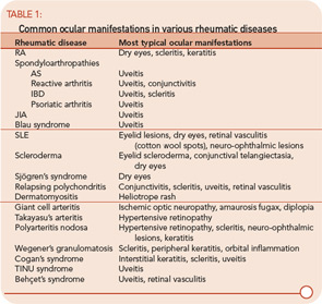
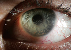
Table 1
click for large version

ad goes here:advert-2
ADVERTISEMENT
SCROLL TO CONTINUE
figure 3: Large concretions of leukocytes adhere to the corneal endothelium (keratic precipitates) in a patient with granulomatous uveitis.

figure 4: Patient with unilateral acute anterior uveitis associated with positive HLA-B27. Right eye shows ciliary injection with irregular pupil, compared to normal left eye.

Ocular involvement is common in rheumatologic disease and varies among different diagnoses. The major ophthalmic manifestations of rheumatic diseases include uveitis, conjunctivitis, scleritis, retinal vasculitis, dry eye syndrome, orbital inflammation, and neuro-ophthalmic lesions. The patterns of common involvement are listed in Table 1 (TK). Among the listed ocular manifestations, uveitis is the most common vision-threatening inflammation associated with rheumatic diseases.1

The etiologies of uveitis include infection, trauma, tumor, or immune-mediated processes. Uveitis is commonly classified by anatomical location of inflammation into anterior, intermediate, posterior, and panuveitis.2

  • Anterior uveitis: When the primary site of inflammation is in the anterior chamber, a synonym for iritis or iridocyclitis.
  • Intermediate uveitis: When the vitreous is the major site of inflammation.
  • Pars planitis: The subset of intermediate uveitis characterized by leukocytic exudates called a snowbank or snowball at the peripheral retina or pars plana.
  • Posterior uveitis: When the retina and/or choroid are the primary site of involvement. Includes including retinitis, choroiditis, chorioretinitis, and retinochoroiditis. The abnormalities can usually be observed by fundoscopic examination.
  • Panuveitis: Reserved for situations in which inflammation is observed in the anterior chamber, vitreous, retina, and/or choroid.
  • Retinal vasculitis: Indicates inflammation in retinal vessels and occasionally occurs without a concomitant uveitis.

Uveitis subsets can also be characterized the following features: onset (sudden versus insidious), laterality (unilateral versus bilateral), duration (limited versus chronic), continuity (recurrent versus persistent), and presence of complications such as the iris sticking to the lens (a phenomenon known as posterior synechiae). Uveitis is termed granulomatous if large concretions of leukocytes adhere to the corneal endothelium (see Figure 3) and non-granulomatous if these concretions are small.

ad goes here:advert-1
ADVERTISEMENT
SCROLL TO CONTINUE

Page: 1 2 3 4 5 6 7 8 9 | Single Page
Share: 

Filed under:Axial SpondyloarthritisBiologics/DMARDsConditionsOther Rheumatic Conditions Tagged with:BiologicsCorticosteroidsDiagnostic Criteriaimmunosuppressive drugsPathogenesisTreatmentUveitis

Related Articles

    An Overview of Pediatric, Noninfectious Uveitis

    October 18, 2018

    Uveitis is an inflammation of the uvea, which comprises the iris, ciliary body and choroid. Uveitis can lead to ocular damage and complete visual loss. Noninfectious etiologies for uveitis are the most common in the U.S.1 The estimated incidence of uveitis ranges from 25–52 per 100,000 in adults and five per 100,000 in children. The…

    ARZTSAMUI / shutterstock.com

    Uveitis: A Brief Primer for the Rheumatologist

    November 12, 2020

    Uveitis is an umbrella term for intraocular inflammatory diseases that can lead to vision loss. It’s not just a concern for ophthalmologists. Uveitis occurs in approximately 2–5% of patients with inflammatory bowel disease, 6–9% of patients with psoriatic arthritis and 25% of patients with reactive arthritis. The prevalence may be as high as 33% in…

    Pediatric Uveitis in the Biologic Age: Risk Factors, Treatment & Outcomes

    March 19, 2019

    CHICAGO—The session on the topic of pediatric uveitis at the 2018 ACR/ARHP Annual Meeting began with a presentation by Debra A. Goldstein, MD, professor of ophthalmology and director of the Uveitis Service at the Feinberg School of Medicine, Northwestern University, Chicago. To a room packed with rheumatologists, she explained, “Most of what I am going…

    Case Report: A Rare Mimic of Giant Cell Arteritis

    November 14, 2021

    Syphilis is a chronic sexually transmitted disease (STD) caused by the spirochete Treponema pallidum. The clinical manifestations of syphilis are divided into four stages: 1) the primary stage, characterized by painless mucosal or cutaneous chancre at the site of infection that resolves spontaneously; 2) the secondary stage in which a generalized maculopapular rash and condyloma…

  • About Us
  • Meet the Editors
  • Issue Archives
  • Contribute
  • Advertise
  • Contact Us
  • Copyright © 2025 by John Wiley & Sons, Inc. All rights reserved, including rights for text and data mining and training of artificial technologies or similar technologies. ISSN 1931-3268 (print). ISSN 1931-3209 (online).
  • DEI Statement
  • Privacy Policy
  • Terms of Use
  • Cookie Preferences