Video: Every Case Tells a Story| Webinar: ACR/CHEST ILD Guidelines in Practice

An official publication of the ACR and the ARP serving rheumatologists and rheumatology professionals

  • Conditions
    • Axial Spondyloarthritis
    • Gout and Crystalline Arthritis
    • Myositis
    • Osteoarthritis and Bone Disorders
    • Pain Syndromes
    • Pediatric Conditions
    • Psoriatic Arthritis
    • Rheumatoid Arthritis
    • Sjögren’s Disease
    • Systemic Lupus Erythematosus
    • Systemic Sclerosis
    • Vasculitis
    • Other Rheumatic Conditions
  • FocusRheum
    • ANCA-Associated Vasculitis
    • Axial Spondyloarthritis
    • Gout
    • Psoriatic Arthritis
    • Rheumatoid Arthritis
    • Systemic Lupus Erythematosus
  • Guidance
    • Clinical Criteria/Guidelines
    • Ethics
    • Legal Updates
    • Legislation & Advocacy
    • Meeting Reports
      • ACR Convergence
      • Other ACR meetings
      • EULAR/Other
    • Research Rheum
  • Drug Updates
    • Analgesics
    • Biologics/DMARDs
  • Practice Support
    • Billing/Coding
    • EMRs
    • Facility
    • Insurance
    • QA/QI
    • Technology
    • Workforce
  • Opinion
    • Patient Perspective
    • Profiles
    • Rheuminations
      • Video
    • Speak Out Rheum
  • Career
    • ACR ExamRheum
    • Awards
    • Career Development
  • ACR
    • ACR Home
    • ACR Convergence
    • ACR Guidelines
    • Journals
      • ACR Open Rheumatology
      • Arthritis & Rheumatology
      • Arthritis Care & Research
    • From the College
    • Events/CME
    • President’s Perspective
  • Search

Case Report: A Patient Presents with Rare, Fulminant SAPHO Syndrome

Ross J. Thibodaux, MD, & Nirupa J. Patel, MD  |  Issue: October 2018  |  October 18, 2018

Tenderness to palpation of his cervical and thoracic spine was noted, and his range of motion in his neck and mid back was severely limited. His right knee was swollen and warm, and his range of motion was decreased due to pain. His left shoulder revealed warmth and swelling, and he had decreased range of motion on adduction and internal and external rotation. A full assessment of his strength was not possible, because pain prevented him from standing up.

Due to the severity and marked constitutional symptoms, he was admitted to the hospital for urgent evaluation. Laboratory analysis revealed elevated C-reactive protein (CRP) of 152.4 mg/L and an erythrocyte sedimentation rate (ESR) of 98 mm/hr. Screening for anti-nuclear antibodies (ANA), rheumatoid factor (RF), human leukocyte antigen B27 (HLA-B27), tuberculosis, human immunodeficiency virus antibodies, viral hepatitis, parvovirus serologies and chlamydial infection was all negative. His cerebrospinal fluid was evaluated for infection, and no evidence of bacterial, fungal or tuberculous meningitis was found.

ad goes here:advert-1
ADVERTISEMENT
SCROLL TO CONTINUE

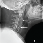
X-rays of his left shoulder revealed mild periosteal elevation and hyperostosis of the proximal humerus and distal acromion, and the radiograph of his right knee revealed a subtle osteolytic, erosive lesion on the lateral side of the femur near the joint margin. Magnetic resonance imaging (MRI) of the cervical, thoracic and lumbar spine revealed multifocal, enhancing lesions consistent with marrow edema (see Figures 2A and 2B, below). The brain MRI did not reveal any intracranial lesions, but clearly revealed the cutaneous abscesses on his scalp (see Figure 2C).
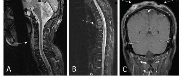
(A) An MRI of the cervical spine revealed multilevel, anterior-predominant, marrow edema extending from C3–C7 with paravertebral edema anteriorly (see white arrow). (B) An MRI of the thoracic spine revealed marrow edema of T3–T5 with mild anterior, superior corner erosion of T5 (see white arrow). (C) An MRI of the brain revealed no intracranial lesions, but note the obvious cellulitis and abscess formation on the scalp (see white arrows).

Figures 2a, b, c
(A) An MRI of the cervical spine revealed multilevel, anterior-predominant, marrow edema extending from C3–C7 with paravertebral edema anteriorly (see white arrow). (B) An MRI of the thoracic spine revealed marrow edema of T3–T5 with mild anterior, superior corner erosion of T5 (see white arrow). (C) An MRI of the brain revealed no intracranial lesions, but note the obvious cellulitis and abscess formation on the scalp (see white arrows).

A biopsy of the T4 vertebral body revealed no evidence of bacterial, acid-fast bacilli, fungal or mycobacterial infection. Surgical pathology of the T4 vertebra revealed only hypercellular marrow tissue without evidence of malignancy.

ad goes here:advert-2
ADVERTISEMENT
SCROLL TO CONTINUE

Presumptive diagnosis: undifferentiated spondyloarthropathy, given his axial and peripheral inflammatory disease. Initial therapy included prednisone and methotrexate.

Clinically, he showed some improvement and was discharged to follow-up care with dermatology and rheumatology clinics. Dermatology performed a biopsy of the scalp lesions that revealed histologic findings consistent with dissecting cellulitis of the scalp. He was also diagnosed with acne keloidalis nuchae.

Page: 1 2 3 4 5 6 | Single Page
Share: 

Filed under:Conditions Tagged with:case reportSAPHO Syndrome

Related Articles

    What Rheumatologists Should Look for in Diagnosing SAPHO Syndrome

    November 1, 2012

    SAPHO syndrome (synovitis, acne, pustulosis, hyperostosis, and osteitis) is an obscure condition that can vex patients and clinicians

    Case Report: Acne Drug Leads Patient to Retinoid Hyperostosis

    November 19, 2018

    Presentation A 26-year-old man with a history of acne vulgaris and hidradenitis suppurativa presented to our rheumatology clinic with persistent back pain and stiffness of three years’ duration. He described bilateral low back pain that was worse when he arose in the morning and at night when he was trying to sleep. In a similar…

    Diagnostic Drill-Down: New Research Suggests Genetic Autoinflammatory Roots

    January 25, 2021

    Two experts discussed ongoing difficulties in diagnosing autoinflammatory disease, & promising new studies that point to possible genetic roots of autoinflammatory disease.

    Tracking Patient Manifestations of Behçet’s Syndrome around the World

    July 1, 2010

    Tracking patient manifestations of Behçet’s syndrome around the world

  • About Us
  • Meet the Editors
  • Issue Archives
  • Contribute
  • Advertise
  • Contact Us
  • Copyright © 2025 by John Wiley & Sons, Inc. All rights reserved, including rights for text and data mining and training of artificial technologies or similar technologies. ISSN 1931-3268 (print). ISSN 1931-3209 (online).
  • DEI Statement
  • Privacy Policy
  • Terms of Use
  • Cookie Preferences