Video: Every Case Tells a Story| Webinar: ACR/CHEST ILD Guidelines in Practice
fa-facebookfa-linkedinfa-youtube-playfa-rss

An official publication of the ACR and the ARP serving rheumatologists and rheumatology professionals

  • Conditions
    • Axial Spondyloarthritis
    • Gout and Crystalline Arthritis
    • Myositis
    • Osteoarthritis and Bone Disorders
    • Pain Syndromes
    • Pediatric Conditions
    • Psoriatic Arthritis
    • Rheumatoid Arthritis
    • Sjögren’s Disease
    • Systemic Lupus Erythematosus
    • Systemic Sclerosis
    • Vasculitis
    • Other Rheumatic Conditions
  • FocusRheum
    • ANCA-Associated Vasculitis
    • Axial Spondyloarthritis
    • Gout
    • Psoriatic Arthritis
    • Rheumatoid Arthritis
    • Systemic Lupus Erythematosus
  • Guidance
    • Clinical Criteria/Guidelines
    • Ethics
    • Legal Updates
    • Legislation & Advocacy
    • Meeting Reports
      • ACR Convergence
      • Other ACR meetings
      • EULAR/Other
    • Research Rheum
  • Drug Updates
    • Analgesics
    • Biologics/DMARDs
  • Practice Support
    • Billing/Coding
    • EMRs
    • Facility
    • Insurance
    • QA/QI
    • Technology
    • Workforce
  • Opinion
    • Patient Perspective
    • Profiles
    • Rheuminations
      • Video
    • Speak Out Rheum
  • Career
    • ACR ExamRheum
    • Awards
    • Career Development
  • ACR
    • ACR Home
    • ACR Convergence
    • ACR Guidelines
    • Journals
      • ACR Open Rheumatology
      • Arthritis & Rheumatology
      • Arthritis Care & Research
    • From the College
    • Events/CME
    • President’s Perspective
  • Search

Case Study in Dermatology: Familial Sarcoidosis/Blau Syndrome

Johanna Sheu, MS, & Joseph F. Merola, MD, MMSc  |  Issue: June 2014  |  June 1, 2014

Multiple erythematous, slightly hyperpigmented waxy papules coalescing into plaques without scale over the bilateral distal lower extremities.
Figure 1: Multiple erythematous, slightly hyperpigmented waxy papules coalescing into plaques without scale over the bilateral distal lower extremities.

ad goes here:advert-1
ADVERTISEMENT
SCROLL TO CONTINUE

The Case

A 43-year-old woman presented with a recurrent rash on her legs. She had a 20-year history of a diagnosis of sarcoidosis, which had been complicated by uveitis and inflammatory arthritis. She also had a history of a subacute granulomatous thyroiditis. Multiple family members also had similar episodes of uveitis, diagnoses of inflammatory arthritis and “sarcoid-like” illnesses.

On examination, she had multiple erythematous, slightly hyperpigmented waxy papules coalescing into plaques without scale over both distal lower extremities (see Figure 1).

ad goes here:advert-2
ADVERTISEMENT
SCROLL TO CONTINUE

What’s Your Diagnosis?

  1. Majocchi’s granuloma/tinea infection
  2. Granuloma annulare
  3. Palisaded neutrophilic granulomatous dermatitis
  4. Familial sarcoidosis/Blau syndrome

Correct answer: D

Familial Sarcoidosis/Blau Syndrome

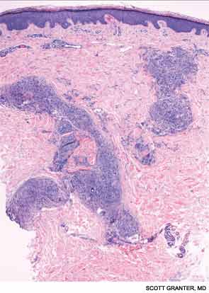
A skin biopsy of a representative lesion was performed, and histopathologic examination revealed nodular aggregates of histiocytes forming non-necrotizing, “naked” granulomas (see Figure 2). Special stains and PCR studies for infectious organisms including mycobacteria were negative. Genetic testing revealed that our patient and multiple family members had a base substitution in the CARD15/NOD2 gene, consistent with a diagnosis of Blau syndrome. Topical steroids were prescribed for her lesions, which resolved with treatment.

Blau syndrome was first described in 1985 by Edward Blau, a pediatrician. It’s a rare, autosomal dominant autoinflammatory disorder with a typical onset within the first two decades of life. Clinically, patients most commonly present with a triad of granulomatous dermatitis, symmetric arthritis and recurrent uveitis. Patients may also have other organ involvement, including hepatic, renal, cardiovascular or neurologic complications.1,2 Mutations that cause Blau syndrome have been mapped to CARD15/NOD2, a caspase recruitment domain gene.

The CARD15/NOD2 gene contains three domains. It is most commonly known for mutations in the third domain, associated with Crohn’s disease, which involves granulomatous inflammation of the bowel. Mutations in the second domain are associated with both Blau syndrome and early onset sarcoidosis. CARD15/NOD2 activates NF-kB in the setting of bacterial cell wall components.3 In Blau syndrome, genetic mutations in the second domain are usually gain-of-function mutations, leading to increased NF-kB activation and possibly increased activity of other inflammatory cytokines.4

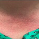
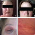
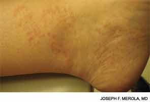
Blau syndrome often presents earliest and most commonly in the skin. Patients present with multiple erythematous and waxy dermal papules and plaques with a lack of scale. Skin biopsy is helpful in diagnosis because it often demonstrates granulomatous inflammation.5

Histopathologic examination revealed nodular aggregates of histiocytes forming non-necrotizing, “naked” granulomas.
Figure 2: Histopathologic examination revealed nodular aggregates of histiocytes forming non-necrotizing, “naked” granulomas.

Discussions of Other Choices

Majocchi’s granuloma occurs when a dermatophyte infection involves the deeper structures of the skin, such as the hair follicles and the dermis. These lesions occur commonly in areas with hair follicles, such as the legs. Patients can also demonstrate other evidence of derma­tophyte infection, such as onychomycosis or tinea pedis. Histopathologic examination of Majocchi’s granuloma would reveal fungal forms on the biopsy.

Granuloma annulare (GA) commonly presents as an erythematous, annular plaque with no scale. However, the subcutaneous form of GA can present as solitary or multiple deeper dermal nodules. Histopathologic examination of these lesions would demonstrate histiocytes around a focus of necrobiosis and increased mucin deposition, the intervening dermis typically appears normal. Multinucleated giant cells are frequently seen along with mild perivascular and interstitial lymphocytic infiltrate with scattered neutrophils.

Palisaded neutrophilic granulomatous dermatitis (PNGD) presents as umbilicated papules distributed symmetrically on the extremities, particularly extensor extremities, as well as on the hands and feet. PNGD is commonly associated with seropositive rheumatoid arthritis and other autoimmune diseases, including systemic lupus erythematosus. On histopathology, PNGD demonstrates a robust neutrophilic and granulomatous histiocytic infiltrate with collagen degeneration and possibly fibrin deposition. These lesions may be thought of as “rheumatoid papules,” a smaller, more superficial variant of the rheumatoid nodule.


Joseph F. Merola, MD, MMSc, is an instructor at Harvard Medical School, Brigham and Women’s Hospital, in Boston. He is board-certified in rheumatology, dermatology and internal medicine. He serves as the director of clinical trials in dermatology, co-director of the Center for Skin and Related Musculoskeletal Diseases and assistant program director for the combined medicine-dermatology residency training program.

Johanna Sheu, MS, is a medical student at Harvard Medical School. She is currently completing a research fellowship in pediatric dermatology at Boston Children’s Hospital.

References

  1. Rose CD, Martin TM, Wouters CH. Blau syndrome revisited. Curr Opin Rheumatol. 2011;23:411–418.
  2. Wang X, Kuivaniemi H, Bonavita G, et al. CARD15 mutations in familial granulomatosis syndromes: A study of the original Blau syndrome kindred and other families with large-vessel arteritis and cranial neuropathy. Arthritis Rheum. 2002;46:3041–3045.
  3. Inohara N, Ogura Y, Fontalba A, et al. Host recognition of bacterial muramyl dipeptide mediated through NOD2. Implications for Crohn’s disease. J Biol Chem. 2003;278:5509–5512.
  4. Sfriso P, Caso F, Tognon S, et al. Blau syndrome, clinical and genetic aspects. Autoimmunity Reviews. 2012;12:44–51.
  5. Janssen CE, Rose CD, De Hertogh G, et al. Morphologic and immunohistochemical characterization of granulomas in the nucleotide oligomerization domain 2-related disorders Blau syndrome and Crohn disease. J Allergy Clin Immunol. 2012;129:1076–1084.

Page: 1 2 | Multi-Page
Share: 

Filed under:ConditionsRheumatoid ArthritisSystemic Lupus ErythematosusVasculitis Tagged with:autoinflammatoryDermatologyRheumatoid arthritisrheumatologistSarcoidosisSystemic lupus erythematosusVasculitis

Related Articles
    Mattew - Bilder und mehr / shutterstock.com

    Yao Syndrome: A Case Report & Clinical Review

    November 12, 2020

    Case Presentation History of present illness A 66-year-old white woman presented with unexplained, recurrent episodes of high fever, abdominal pain, rash and arthralgias occurring over the previous three years. During typical episodes, the patient experienced flu-like symptoms, followed by fever, abdominal pain and non-bloody diarrhea without tenesmus. ad goes here:advert-1ADVERTISEMENTSCROLL TO CONTINUEHer temperatures were 101–103ºF,…

    Case Report: A Long, Arduous Evaluation Capped by Genetic Testing

    February 14, 2023

    FIGURE 1: Cutaneous flushing and patchy erythematous macules on the neck and upper chest. Systemic autoinflammatory diseases (SAIDs) are rare syndromes characterized by alterations in innate immunity that result in a variety of clinical manifestations that are usually associated with recurrent fevers.1 Thanks to advances in genetic sequencing over the past few years, monogenic causes…

    The Granulomatosis of Wegener’s

    May 16, 2011

    Delving deeper into the nonvasculitis aspects of the disease

    Fellows’ Forum Case Report: Autoinflammatory Diseases and Roles of Genetic, Molecular Testing

    October 17, 2017

    Figure 1. Patient’s rash. A and B: swollen and erythematous patches with maculopapular changes on the face; C: eyelid swelling with discoloration; D: erythematous patches and plaques on abdominal wall. A 51-year-old Caucasian female was referred by a local rheumatologist to the Center of Autoinflammatory Diseases at Stony Brook University, N.Y., for an unusual disease…

  • About Us
  • Meet the Editors
  • Issue Archives
  • Contribute
  • Advertise
  • Contact Us
fa-facebookfa-linkedinfa-youtube-playfa-rss
  • Copyright © 2025 by John Wiley & Sons, Inc. All rights reserved, including rights for text and data mining and training of artificial technologies or similar technologies. ISSN 1931-3268 (print). ISSN 1931-3209 (online).
  • DEI Statement
  • Privacy Policy
  • Terms of Use
  • Cookie Preferences