Amyloid arthropathy is a known complication of long-term hemodialysis, characterized by the intraosseous, intra-articular and periarticular deposition of a unique form of amyloid composed of beta-2-microglobulin. This amyloid deposition in renal failure patients has a propensity for the musculoskeletal system, and the prevalence increases with the duration of dialysis therapy. The articular lesions are characterized by soft tissue masses, well-defined periarticular erosions and relative preservation of the joint space.
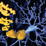
Figure 1.
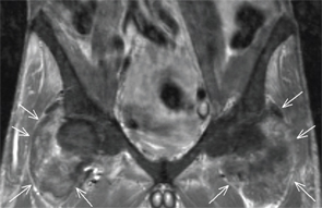
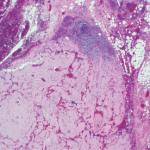
Figure 2.
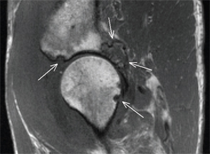
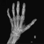
Figure 3.
Jennifer L. Demertzis, MD, is a musculoskeletal radiologist at the Mallinckrodt Institute of Radiology at Washington University School of Medicine in St. Louis, Mo. She is excited to collaborate on this new feature in the journal and looks forward to seeing future cases contributed by readers.