The Rheumatologist
COVID-19 NewsACR Convergence
  • Connect with us:
  • Facebook
  • Twitter
  • LinkedIn
  • YouTube
  • Feed
  • Home
  • Conditions
    • Rheumatoid Arthritis
    • SLE (Lupus)
    • Crystal Arthritis
      • Gout Resource Center
    • Spondyloarthritis
    • Osteoarthritis
    • Soft Tissue Pain
    • Scleroderma
    • Vasculitis
    • Systemic Inflammatory Syndromes
    • Guidelines
  • Resource Centers
    • Axial Spondyloarthritis Resource Center
    • Gout Resource Center
    • Psoriatic Arthritis Resource Center
    • Rheumatoid Arthritis Resource Center
    • Systemic Lupus Erythematosus Resource Center
  • Drug Updates
    • Biologics & Biosimilars
    • DMARDs & Immunosuppressives
    • Topical Drugs
    • Analgesics
    • Safety
    • Pharma Co. News
  • Professional Topics
    • Ethics
    • Legal
    • Legislation & Advocacy
    • Career Development
      • Certification
      • Education & Training
    • Awards
    • Profiles
    • President’s Perspective
    • Rheuminations
    • Interprofessional Perspective
  • Practice Management
    • Billing/Coding
    • Quality Assurance/Improvement
    • Workforce
    • Facility
    • Patient Perspective
    • Electronic Health Records
    • Apps
    • Information Technology
    • From the College
    • Multimedia
      • Audio
      • Video
  • Resources
    • Issue Archives
    • ACR Convergence
      • Gout Resource Center
      • Axial Spondyloarthritis Resource Center
      • Psoriatic Arthritis
      • Abstracts
      • Meeting Reports
      • ACR Convergence Home
    • American College of Rheumatology
    • ACR ExamRheum
    • Research Reviews
    • ACR Journals
      • Arthritis & Rheumatology
      • Arthritis Care & Research
      • ACR Open Rheumatology
    • Rheumatology Image Library
    • Treatment Guidelines
    • Rheumatology Research Foundation
    • Events
  • About Us
    • Mission/Vision
    • Meet the Authors
    • Meet the Editors
    • Contribute to The Rheumatologist
    • Subscription
    • Contact
  • Advertise
  • Search
You are here: Home / Articles / Diagnostic Imaging in Patient with Left Hip Pain: Findings

Diagnostic Imaging in Patient with Left Hip Pain: Findings

August 18, 2015 • By Jennifer L. Demertzis, MD

  • Tweet
  • Email
Print-Friendly Version / Save PDF

View the question.

You Might Also Like
  • Diagnostic Imaging in Patient with Left Hip Pain: History
  • Diagnostic Imaging in Patient with Atraumatic Left Shoulder Pain: Findings
  • Diagnostic Imaging in Patient with Chronic Left Ankle Pain: Findings
Explore This Issue
August 2015
Also By This Author
  • Diagnostic Imaging in Patient with Soft Tissue Calcification: Findings

Findings/Diagnosis

The AP radiograph of the left hip (see Figure 1) shows periarticular, well-defined erosions of the left hip (white arrow) without joint space narrowing or osteophytes. There is no fracture. There are surgical clips and a calcified mass in the right hemipelvis (black ellipsis), representing a failed renal transplant.

ad goes here:advert-1
ADVERTISEMENT
SCROLL TO CONTINUE

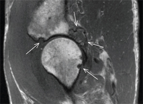
The coronal STIR image of the pelvis demonstrates hypointense intra-articular synovial masses in the right and left hips (see Figure 2, white arrows). There are multiple synovial pressure erosions in the left acetabulum and femur (see Figure 3, white arrows); the articular cartilage otherwise appears preserved.

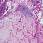
Based on these imaging findings and the history of chronic hemodialysis, the differential diagnosis included gout and amyloidosis. Given the polyarticular nature, pigmented villonodular synovitis was considered less likely. An ultrasound-guided synovial biopsy of the left hip was performed. Gram stain, culture and crystal analysis were negative. Surgical pathology revealed synovial tissue with nodular masses of extracellular, amorphous, eosinophilic material that was Congo-red positive, consistent with amyloid deposition. The patient had an elevated serum beta-2-microglobulin (25.8 mg/L; normal range 0.7–3.4 mg/L), further supporting the diagnosis.

ad goes here:advert-2
ADVERTISEMENT
SCROLL TO CONTINUE

Amyloid arthropathy is a known complication of long-term hemodialysis, characterized by the intraosseous, intra-articular and periarticular deposition of a unique form of amyloid composed of beta-2-microglobulin. This amyloid deposition in renal failure patients has a propensity for the musculoskeletal system, and the prevalence increases with the duration of dialysis therapy. The articular lesions are characterized by soft tissue masses, well-defined periarticular erosions and relative preservation of the joint space.
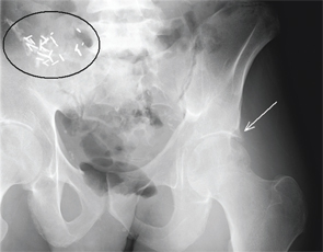
Figure 1.
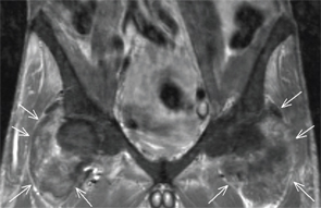
Figure 1.

Figure 2.

Figure 2.

Figure 3.

Figure 3.


Jennifer L. Demertzis, MD, is a musculoskeletal radiologist at the Mallinckrodt Institute of Radiology at Washington University School of Medicine in St. Louis, Mo. She is excited to collaborate on this new feature in the journal and looks forward to seeing future cases contributed by readers.
ad goes here:advert-3
ADVERTISEMENT
SCROLL TO CONTINUE

Filed Under: Conditions, Soft Tissue Pain Tagged With: Diagnosis, diagnostic imaging, hip pain, imaging, MRI, patient care, radiographIssue: August 2015

You Might Also Like:
  • Diagnostic Imaging in Patient with Left Hip Pain: History
  • Diagnostic Imaging in Patient with Atraumatic Left Shoulder Pain: Findings
  • Diagnostic Imaging in Patient with Chronic Left Ankle Pain: Findings
  • Diagnostic Imaging in Patient with Soft Tissue Calcification: Findings

Simple Tasks

Learn more about the ACR’s public awareness campaign and how you can get involved. Help increase visibility of rheumatic diseases and decrease the number of people left untreated.

Visit the Simple Tasks site »

American College of Rheumatology

Visit the official website for the American College of Rheumatology.

Visit the ACR »

ACR Convergence

Don’t miss rheumatology’s premier scientific meeting for anyone involved in research or the delivery of rheumatologic care or services.

Visit the ACR Convergence site »

The Rheumatologist newsmagazine reports on issues and trends in the management and treatment of rheumatic diseases. The Rheumatologist reaches 11,500 rheumatologists, internists, orthopedic surgeons, nurse practitioners, physician assistants, nurses, and other healthcare professionals who practice, research, or teach in the field of rheumatology.

About Us / Contact Us / Advertise / Privacy Policy / Terms of Use / Cookie Preferences

  • Connect with us:
  • Facebook
  • Twitter
  • LinkedIn
  • YouTube
  • Feed

Copyright © 2006–2023 American College of Rheumatology. All rights reserved.

ISSN 1931-3268 (print)
ISSN 1931-3209 (online)