Video: Every Case Tells a Story| Webinar: ACR/CHEST ILD Guidelines in Practice

An official publication of the ACR and the ARP serving rheumatologists and rheumatology professionals

  • Conditions
    • Axial Spondyloarthritis
    • Gout and Crystalline Arthritis
    • Myositis
    • Osteoarthritis and Bone Disorders
    • Pain Syndromes
    • Pediatric Conditions
    • Psoriatic Arthritis
    • Rheumatoid Arthritis
    • Sjögren’s Disease
    • Systemic Lupus Erythematosus
    • Systemic Sclerosis
    • Vasculitis
    • Other Rheumatic Conditions
  • FocusRheum
    • ANCA-Associated Vasculitis
    • Axial Spondyloarthritis
    • Gout
    • Psoriatic Arthritis
    • Rheumatoid Arthritis
    • Systemic Lupus Erythematosus
  • Guidance
    • Clinical Criteria/Guidelines
    • Ethics
    • Legal Updates
    • Legislation & Advocacy
    • Meeting Reports
      • ACR Convergence
      • Other ACR meetings
      • EULAR/Other
    • Research Rheum
  • Drug Updates
    • Analgesics
    • Biologics/DMARDs
  • Practice Support
    • Billing/Coding
    • EMRs
    • Facility
    • Insurance
    • QA/QI
    • Technology
    • Workforce
  • Opinion
    • Patient Perspective
    • Profiles
    • Rheuminations
      • Video
    • Speak Out Rheum
  • Career
    • ACR ExamRheum
    • Awards
    • Career Development
  • ACR
    • ACR Home
    • ACR Convergence
    • ACR Guidelines
    • Journals
      • ACR Open Rheumatology
      • Arthritis & Rheumatology
      • Arthritis Care & Research
    • From the College
    • Events/CME
    • President’s Perspective
  • Search

Fellow’s Forum Case Report: Takayasu’s Arteritis

Ashwini Komarla, MD; Michael George, MD; Antoine Sreih, MD; & Chris Derk, MD, MS  |  Issue: May 2014  |  May 1, 2014

Fellow's Forum: Takayasu’s Arteritis

ad goes here:advert-1
ADVERTISEMENT
SCROLL TO CONTINUE

Case 1

A 27-year-old Caucasian woman previously in good health presented to the emergency department (ED) with three months of progressive fatigue, shortness of breath, paroxysmal nocturnal dyspnea and orthopnea. She also described left flank pain, left frontal neck and upper back pain, left arm claudication and frequent left-side headaches. She had an episode of complete loss of vision in her left eye lasting a few seconds three months prior to admission, but she did not seek medical attention at the time. Her progressive shortness of breath prompted her ED visit.

A chest computed tomography (CT) scan was performed to evaluate for a pulmonary embolus, but instead it revealed dilation of the ascending aorta and a 3.9 cm wide fusiform aneurysm of the descending thoracic aorta. Her blood pressure was 98/49 mmHg in her right arm, 88/36 mmHg in her left arm, 133/26 and 135/26 mmHg in her right and left legs. Her physical exam was significant for a IV/VI diastolic murmur loudest at the right upper sternal border, a barely palpable left carotid pulse, and weak left axillary, brachial and radial pulses. The rest of her vascular exam was normal.

ad goes here:advert-2
ADVERTISEMENT
SCROLL TO CONTINUE

Her basic laboratory tests were unremarkable except for an anemia with a hemoglobin of 11.9 (reference 12.0–16.0 g/dL). A high-sensitivity C-reactive protein (hs-CRP) was 19 (reference <7.4 mg/L) with an erythrocyte sedimentation rate (ESR) of 5 (reference <25 mm/hr). A transesophageal echocardiogram revealed thickening of the ascending aorta, severe aortic insufficiency with a flail right cusp, aortic root dilation and thickening of the valve. Her left ventricular ejection fraction was 35%.

CT angiography of the neck axial view demonstrating complete occlusion of left common carotid artery (white arrow) and significant wall thickening of the left subclavian artery (red arrow)
Figure 1: CT angiography of the neck axial view demonstrating complete occlusion of left common carotid artery (white arrow) and significant wall thickening of the left subclavian artery (red arrow).

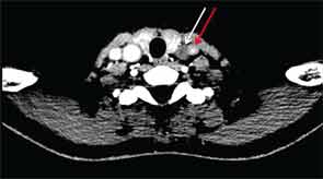
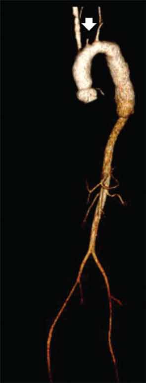
CT of the neck showed complete occlusion of left common carotid artery immediately above the stump (see Figure 1) and complete occlusion of the left internal carotid artery with retrograde filling via the circle of Willis. Significant wall thickening and multiple areas of irregularity and dilatation were seen in the right innominate, right common carotid and left subclavian arteries. A CT angiogram (CTA) of the chest, abdomen and pelvis demonstrated a dilated ascending and descending thoracic aorta with a maximum dimension of 4 cm with concentric wall thickening extending to the midthoracic aorta (see Figure 2). There was no involvement of her abdominal aorta.

CT angiogram of chest, abdomen and pelvis
Figure 2: CT angiogram of chest, abdomen and pelvis with 3D reconstruction of the aortic arch and major vessels. There is dilatation and mural thickening of the ascending and descending thoracic aorta with a maximum outer diameter of 4 cm that extends to the midthoracic aorta above the diaphragmatic hiatus. There is redemonstration of the complete occlusion of the left common carotid artery (white arrow). The abdominal aorta, celiac plexus, renal and iliac arteries are unremarkable.

The patient was given a pulse dose of glucocorticoids with methylprednisolone 500 mg intravenous daily for three days with improvement in the strength of her left brachial and radial pulses over the course of treatment. She was also initiated on aspirin 81 mg daily and azathioprine 50 mg daily, which was further titrated as an outpatient. After the glucocorticoid pulse, the dose was reduced to prednisone 1 mg/kg/day. Aortic valve replacement was deferred in order to treat the inflammatory vascular process and because she was well compensated clinically. She returned a month later and continued to have stable heart failure symptoms, although she had episodes of left-side amaurosis fugax, lasting less than 15 seconds. Her glucocorticoid dose will be tapered in anticipation of an aortic valve replacement.

Case 2

A 31-year-old Hispanic woman presented to the hospital with hypertension and renal failure.

Page: 1 2 3 4 5 6 7 | Single Page
Share: 

Filed under:ConditionsVasculitis Tagged with:drugGiant Cell ArteritisGlucocorticoidsimaginginfliximablarge-vessel vasculitisMethotrexatePainpatient careprednisonerheumatologistrituximabTakayasu’s ArteritistocilizumabTreatmentVasculitis

Related Articles

    Fellow’s Forum Case Report: Aneurysm in Takayasu’s Arteritis

    August 17, 2015

    A healthy 30-year-old Indian male was performing his routine workout in the gym when he developed giddiness and transient blackouts. He thought it was due to exercising excessively. Over the next month, he developed a low-grade fever and started feeling lethargic, to the extent that he was unable to exercise or work. He was admitted…

    Study Finds Tocilizumab Could Be Treatment Option for Takayasu Arteritis

    August 17, 2018

    For patients with refractory Takayasu arteritis (TAK), glucocorticoids (GCs) are often provided as the initial therapy for treatment. However, GCs are often associated with adverse effects for long-term use; relapse also occurs frequently during GC tapering.1 TAK involves interleukin (IL) 6. Tocilizumab—a recombinant, humanized, anti-IL-6 receptor (IL-6R) monoclonal antibody—was first reported by Nishimoto et al….

    Patients with Large-Vessel Abnormalities Present Diagnostic Dilemma for Rheumatologists

    February 1, 2015

    Absent signs, symptoms suggesting etiology, clinicians rely on inflammatory markers, imaging to make the diagnosis

    Two Inflammatory Conditions—Polymyalgia Rheumatica and Giant Cell Arteritis—Share Clinical Connection

    March 1, 2013

    Polymyalgia rheumatica (PMR) and giant cell arteritis (GCA) have common clinical and epidemiologic links, but they need not occur synchronously

  • About Us
  • Meet the Editors
  • Issue Archives
  • Contribute
  • Advertise
  • Contact Us
  • Copyright © 2025 by John Wiley & Sons, Inc. All rights reserved, including rights for text and data mining and training of artificial technologies or similar technologies. ISSN 1931-3268 (print). ISSN 1931-3209 (online).
  • DEI Statement
  • Privacy Policy
  • Terms of Use
  • Cookie Preferences