Video: Every Case Tells a Story| Webinar: ACR/CHEST ILD Guidelines in Practice

An official publication of the ACR and the ARP serving rheumatologists and rheumatology professionals

  • Conditions
    • Axial Spondyloarthritis
    • Gout and Crystalline Arthritis
    • Myositis
    • Osteoarthritis and Bone Disorders
    • Pain Syndromes
    • Pediatric Conditions
    • Psoriatic Arthritis
    • Rheumatoid Arthritis
    • Sjögren’s Disease
    • Systemic Lupus Erythematosus
    • Systemic Sclerosis
    • Vasculitis
    • Other Rheumatic Conditions
  • FocusRheum
    • ANCA-Associated Vasculitis
    • Axial Spondyloarthritis
    • Gout
    • Psoriatic Arthritis
    • Rheumatoid Arthritis
    • Systemic Lupus Erythematosus
  • Guidance
    • Clinical Criteria/Guidelines
    • Ethics
    • Legal Updates
    • Legislation & Advocacy
    • Meeting Reports
      • ACR Convergence
      • Other ACR meetings
      • EULAR/Other
    • Research Rheum
  • Drug Updates
    • Analgesics
    • Biologics/DMARDs
  • Practice Support
    • Billing/Coding
    • EMRs
    • Facility
    • Insurance
    • QA/QI
    • Technology
    • Workforce
  • Opinion
    • Patient Perspective
    • Profiles
    • Rheuminations
      • Video
    • Speak Out Rheum
  • Career
    • ACR ExamRheum
    • Awards
    • Career Development
  • ACR
    • ACR Home
    • ACR Convergence
    • ACR Guidelines
    • Journals
      • ACR Open Rheumatology
      • Arthritis & Rheumatology
      • Arthritis Care & Research
    • From the College
    • Events/CME
    • President’s Perspective
  • Search

Fellow’s Forum Case Report: Takayasu’s Arteritis

Ashwini Komarla, MD; Michael George, MD; Antoine Sreih, MD; & Chris Derk, MD, MS  |  Issue: May 2014  |  May 1, 2014

The American College of Rheumatology has established classification criteria for TAK (see Table 1), which were primarily designed to distinguish this disorder from other forms of vasculitis for study purposes. This classification system has a sensitivity and specificity of 90.5% and 97.8%, respectively.6 Attempts have been made to classify the disease on the basis of angiographic findings (see Figure 4), which can be useful in allowing a comparison of patient characteristics according to the involved vessels and helpful in planning surgery.7 However, this provides little information on prognosis.

ad goes here:advert-1
ADVERTISEMENT
SCROLL TO CONTINUE
Angiographic classification of Takayasu’s arteritis
click for large version
Figure 4: Angiographic classification of Takayasu’s arteritis from the Takayasu conference in 1994.
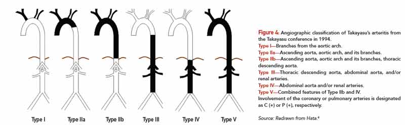
Type I—Branches from the aortic arch.
Type IIa—Ascending aorta, aortic arch, and its branches.
Type IIb—Ascending aorta, aortic arch and its branches, thoracic descending aorta.
Type III—Thoracic descending aorta, abdominal aorta, and/or renal arteries.
Type IV—Abdominal aorta and/or renal arteries.
Type V—Combined features of Type IIb and IV.
Involvement of the coronary or pulmonary arteries is designated as C (+) or P (+), respectively.
Source: Redrawn from Hata.6

Conventional angiography remains the gold standard for the diagnosis of TAK. MRA and CTA can also be used to evaluate large- and medium-size vessels. Rheumatic diseases that affect the aorta or its branches include giant cell arteritis, primarily differentiated from Takayasu’s by age of disease onset and distribution of lesions. Granulomatous vasculitis can be demonstrated on histologic examination in both diseases. Other rheumatic diseases, such as relapsing polychondritis, ankylosing spondylitis, Behçet’s disease and Cogan’s syndrome, may have a similar arteriographic appearance to TAK but can be differentiated on the basis of their other clinical manifestations. Infectious or “mycotic” aneurysms, atherosclerotic disease, fibromuscular dysplasia, congenital co-arctation and inherited disorders of connective tissue, such as Marfan’s syndrome and Ehlers Danlos, must also be considered.

The mainstay of therapy is suppression of inflammation and preservation of vascular competence. Initial treatment is typically glucocorticoids at a dose of 1 mg/kg/day. Although the majority of patients will respond to this treatment, some cases are glucocorticoid resistant. In addition, relapse with tapering steroids is common. Most patients will require other immunosuppressive agents, which are often initiated at the time of diagnosis.8 An initial open-label study of methotrexate with glucocorticoids in 16 patients showed remission in 81% of patients and sustained remission in 50% of patients, suggesting its utility as a steroid-sparing agent.9 Limited data also exists to support the use of azathioprine, mycophenolate, cyclosporine, tacrolimus, leflunomide or cyclophosphamide.10 In all cases, a significant number of patients continue to have active disease or are unable to discontinue glucocorticoid treatment.

ad goes here:advert-2
ADVERTISEMENT
SCROLL TO CONTINUE

Page: 1 2 3 4 5 6 7 | Single Page
Share: 

Filed under:ConditionsVasculitis Tagged with:drugGiant Cell ArteritisGlucocorticoidsimaginginfliximablarge-vessel vasculitisMethotrexatePainpatient careprednisonerheumatologistrituximabTakayasu’s ArteritistocilizumabTreatmentVasculitis

Related Articles

    Fellow’s Forum Case Report: Aneurysm in Takayasu’s Arteritis

    August 17, 2015

    A healthy 30-year-old Indian male was performing his routine workout in the gym when he developed giddiness and transient blackouts. He thought it was due to exercising excessively. Over the next month, he developed a low-grade fever and started feeling lethargic, to the extent that he was unable to exercise or work. He was admitted…

    Study Finds Tocilizumab Could Be Treatment Option for Takayasu Arteritis

    August 17, 2018

    For patients with refractory Takayasu arteritis (TAK), glucocorticoids (GCs) are often provided as the initial therapy for treatment. However, GCs are often associated with adverse effects for long-term use; relapse also occurs frequently during GC tapering.1 TAK involves interleukin (IL) 6. Tocilizumab—a recombinant, humanized, anti-IL-6 receptor (IL-6R) monoclonal antibody—was first reported by Nishimoto et al….

    Patients with Large-Vessel Abnormalities Present Diagnostic Dilemma for Rheumatologists

    February 1, 2015

    Absent signs, symptoms suggesting etiology, clinicians rely on inflammatory markers, imaging to make the diagnosis

    Two Inflammatory Conditions—Polymyalgia Rheumatica and Giant Cell Arteritis—Share Clinical Connection

    March 1, 2013

    Polymyalgia rheumatica (PMR) and giant cell arteritis (GCA) have common clinical and epidemiologic links, but they need not occur synchronously

  • About Us
  • Meet the Editors
  • Issue Archives
  • Contribute
  • Advertise
  • Contact Us
  • Copyright © 2025 by John Wiley & Sons, Inc. All rights reserved, including rights for text and data mining and training of artificial technologies or similar technologies. ISSN 1931-3268 (print). ISSN 1931-3209 (online).
  • DEI Statement
  • Privacy Policy
  • Terms of Use
  • Cookie Preferences