Video: Every Case Tells a Story| Webinar: ACR/CHEST ILD Guidelines in Practice

An official publication of the ACR and the ARP serving rheumatologists and rheumatology professionals

  • Conditions
    • Axial Spondyloarthritis
    • Gout and Crystalline Arthritis
    • Myositis
    • Osteoarthritis and Bone Disorders
    • Pain Syndromes
    • Pediatric Conditions
    • Psoriatic Arthritis
    • Rheumatoid Arthritis
    • Sjögren’s Disease
    • Systemic Lupus Erythematosus
    • Systemic Sclerosis
    • Vasculitis
    • Other Rheumatic Conditions
  • FocusRheum
    • ANCA-Associated Vasculitis
    • Axial Spondyloarthritis
    • Gout
    • Psoriatic Arthritis
    • Rheumatoid Arthritis
    • Systemic Lupus Erythematosus
  • Guidance
    • Clinical Criteria/Guidelines
    • Ethics
    • Legal Updates
    • Legislation & Advocacy
    • Meeting Reports
      • ACR Convergence
      • Other ACR meetings
      • EULAR/Other
    • Research Rheum
  • Drug Updates
    • Analgesics
    • Biologics/DMARDs
  • Practice Support
    • Billing/Coding
    • EMRs
    • Facility
    • Insurance
    • QA/QI
    • Technology
    • Workforce
  • Opinion
    • Patient Perspective
    • Profiles
    • Rheuminations
      • Video
    • Speak Out Rheum
  • Career
    • ACR ExamRheum
    • Awards
    • Career Development
  • ACR
    • ACR Home
    • ACR Convergence
    • ACR Guidelines
    • Journals
      • ACR Open Rheumatology
      • Arthritis & Rheumatology
      • Arthritis Care & Research
    • From the College
    • Events/CME
    • President’s Perspective
  • Search

Metagenomic Deep Sequencing for Uveitis Enhances Traditional Diagnostic Testing

Thuy Doan, MD, PhD, Michael R. Wilson, MD, MAS, & Joseph L. DeRisi, PhD  |  Issue: March 2017  |  March 20, 2017

The relative inaccessibility of the intraocular compartments (i.e., anterior chamber and vitreous cavity) poses an obvious limitation to performing an extensive diagnostic work-up, as does the minute volumes of fluid that are available even if a diagnostic paracentesis is performed. Even the most advanced centers offer only a handful of microbiologic assays for intraocular fluid including culture (e.g., bacterial, acid fast bacilli, fungal and rarely viral) and pathogen-specific polymerase chain reactions (e.g., herpes simplex virus types 1 and 2, varicella zoster virus, cytomegalovirus, Toxoplasma gondii).

Not surprisingly, an etiologic agent is not identified in more than half of uveitis cases, and physicians are forced to make empiric treatment decisions. If local or systemic immunosuppression is pursued to treat a presumed autoimmune cause of uveitis, this can serve to exacerbate an undiagnosed intraocular infection.

ad goes here:advert-1
ADVERTISEMENT
SCROLL TO CONTINUE

We first used MDS in three uveitis cases in which a pathogen had already been identified and showed that MDS was consistent with the gold standard microbiologic testing for herpes-simplex virus type 1, Cryptococcus neoformans and Toxoplasma gondii.1

We also showed that in two noninfectious intraocular fluid samples, MDS did not detect any pathogenic organisms. Rather, the MDS results from these two samples largely reflected organisms that are found in laboratory reagents.

ad goes here:advert-2
ADVERTISEMENT
SCROLL TO CONTINUE

Case Study

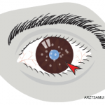
After showing that MDS could be used to identify common infectious causes of uveitis, we turned to a patient with a 16-year course of chronic, idiopathic, asynchronous bilateral uveitis. In 1999, he noticed floaters and mild decreased vision in the left eye. The patient was seen by an ophthalmologist in Germany and found to have anterior uveitis, which is the most common uveitis entity.

A routine laboratory work-up (e.g., HLA-B27, RF, c-ANCA, hepatitis serology panel and PPD) was unrevealing. The patient’s intraocular inflammation was managed with topical steroids and non-steroidal anti-inflammatory drugs (NSAIDs) with minimal improvement.

Two years later, the patient had ocular symptoms in the right eye and was diagnosed with bilateral idiopathic anterior uveitis. RPR and repeat HLA-B27 were negative. At this time, he had already had cataract surgery in the left eye, likely due to a combination of topical steroid and uncontrolled intraocular inflammation. Seven years after disease onset, the patient was initiated on a potent topical steroid (Durezol or difluprednate). That failed to control his inflammation, so he was placed on systemic prednisone and methotrexate, in addition to frequent topical steroid drops.

Page: 1 2 3 4 5 | Single Page
Share: 

Filed under:ConditionsOther Rheumatic Conditions Tagged with:diagnostichypothesis-freeinflammatory syndromeintraocular infectionmetagenomic deep sequencingpatient carerheumatologistrheumatologyTestUveitis

Related Articles

    An Overview of Pediatric, Noninfectious Uveitis

    October 18, 2018

    Uveitis is an inflammation of the uvea, which comprises the iris, ciliary body and choroid. Uveitis can lead to ocular damage and complete visual loss. Noninfectious etiologies for uveitis are the most common in the U.S.1 The estimated incidence of uveitis ranges from 25–52 per 100,000 in adults and five per 100,000 in children. The…

    Watch Those Eyes

    December 1, 2007

    What you need to know about Uveitis in Rheumatic Diseases

    ARZTSAMUI / shutterstock.com

    Uveitis: A Brief Primer for the Rheumatologist

    November 12, 2020

    Uveitis is an umbrella term for intraocular inflammatory diseases that can lead to vision loss. It’s not just a concern for ophthalmologists. Uveitis occurs in approximately 2–5% of patients with inflammatory bowel disease, 6–9% of patients with psoriatic arthritis and 25% of patients with reactive arthritis. The prevalence may be as high as 33% in…

    Pediatric Uveitis in the Biologic Age: Risk Factors, Treatment & Outcomes

    March 19, 2019

    CHICAGO—The session on the topic of pediatric uveitis at the 2018 ACR/ARHP Annual Meeting began with a presentation by Debra A. Goldstein, MD, professor of ophthalmology and director of the Uveitis Service at the Feinberg School of Medicine, Northwestern University, Chicago. To a room packed with rheumatologists, she explained, “Most of what I am going…

  • About Us
  • Meet the Editors
  • Issue Archives
  • Contribute
  • Advertise
  • Contact Us
  • Copyright © 2025 by John Wiley & Sons, Inc. All rights reserved, including rights for text and data mining and training of artificial technologies or similar technologies. ISSN 1931-3268 (print). ISSN 1931-3209 (online).
  • DEI Statement
  • Privacy Policy
  • Terms of Use
  • Cookie Preferences