Video: Every Case Tells a Story| Webinar: ACR/CHEST ILD Guidelines in Practice

An official publication of the ACR and the ARP serving rheumatologists and rheumatology professionals

  • Conditions
    • Axial Spondyloarthritis
    • Gout and Crystalline Arthritis
    • Myositis
    • Osteoarthritis and Bone Disorders
    • Pain Syndromes
    • Pediatric Conditions
    • Psoriatic Arthritis
    • Rheumatoid Arthritis
    • Sjögren’s Disease
    • Systemic Lupus Erythematosus
    • Systemic Sclerosis
    • Vasculitis
    • Other Rheumatic Conditions
  • FocusRheum
    • ANCA-Associated Vasculitis
    • Axial Spondyloarthritis
    • Gout
    • Psoriatic Arthritis
    • Rheumatoid Arthritis
    • Systemic Lupus Erythematosus
  • Guidance
    • Clinical Criteria/Guidelines
    • Ethics
    • Legal Updates
    • Legislation & Advocacy
    • Meeting Reports
      • ACR Convergence
      • Other ACR meetings
      • EULAR/Other
    • Research Rheum
  • Drug Updates
    • Analgesics
    • Biologics/DMARDs
  • Practice Support
    • Billing/Coding
    • EMRs
    • Facility
    • Insurance
    • QA/QI
    • Technology
    • Workforce
  • Opinion
    • Patient Perspective
    • Profiles
    • Rheuminations
      • Video
    • Speak Out Rheum
  • Career
    • ACR ExamRheum
    • Awards
    • Career Development
  • ACR
    • ACR Home
    • ACR Convergence
    • ACR Guidelines
    • Journals
      • ACR Open Rheumatology
      • Arthritis & Rheumatology
      • Arthritis Care & Research
    • From the College
    • Events/CME
    • President’s Perspective
  • Search

Metagenomic Deep Sequencing for Uveitis Enhances Traditional Diagnostic Testing

Thuy Doan, MD, PhD, Michael R. Wilson, MD, MAS, & Joseph L. DeRisi, PhD  |  Issue: March 2017  |  March 20, 2017

He eventually developed a white cataract in the right eye and underwent cataract surgery and IOL placement. His immunosuppression was discontinued after one year due to treatment futility. There was a discussion to transition the patient to another antimetabolite, CellCept, but he never started the medication due to recurrent lower extremity rashes.

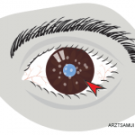
The patient relocated to San Francisco in 2012 and sought care at the Proctor Foundation at the University of California, San Francisco. His exam was notable for inflammatory cells in the anterior and vitreous cavity bilaterally. Further, he had heterochromia (different iris color between the two eyes) suggestive of chronic viral infection.

ad goes here:advert-1
ADVERTISEMENT
SCROLL TO CONTINUE

An anterior chamber paracentesis was performed, and aqueous fluid was tested for HSV, VZV and CMV by polymerase chain reaction (PCR). Although the results were negative for herpesvirus infection, the patient was maintained on systemic and intravitreal antivirals because of the high degree of clinical suspicion. When he had to undergo a pars plana vitrectomy to remove an epiretinal membrane in the left eye, another diagnostic work-up was initiated, which included cytology, bacterial and fungal cultures, HSV, VZV and CMV PCRs. Again, all tests were nondiagnostic.

The patient’s aqueous fluid was finally evaluated by unbiased MDS. After filtering out host sequencing reads and environmental reads, the most predominant remaining viral reads aligned to rubella virus, a known infectious cause of uveitis. More importantly, a near full-length genome (99.3%) of the rubella virus was obtained. Phylogenetic analysis revealed that the rubella virus detected in the patient’s eye was mostly closely related to a rubella virus isolated in Stuttgart, Germany, in 1992.

ad goes here:advert-2
ADVERTISEMENT
SCROLL TO CONTINUE

Upon further questioning, the patient reported that he had had a three-day fever and whole body rash in 1993 when he was living in Germany. Because of the German vaccination policies at the time, he had not been immunized against rubella virus. Thus, it appears as if his ocular history actually began in 1993, a full six years prior to the onset of his uveitis.

In this case, MDS not only identified a very unusual cause of infectious uveitis, it also highlighted the Achilles heel of pattern recognition as a primary means of diagnosis in patients with inflammatory disease. In the vast majority of cases, a patient’s three-day fever and rash that had occurred six years prior to the onset of their current clinical syndrome would not be considered clinically relevant—or even asked about by most clinicians. Thus, MDS has the ability to not only identify unusual pathogens, but it will also help redefine clinical syndromes by its unbiased and comprehensive approach.

Page: 1 2 3 4 5 | Single Page
Share: 

Filed under:ConditionsOther Rheumatic Conditions Tagged with:diagnostichypothesis-freeinflammatory syndromeintraocular infectionmetagenomic deep sequencingpatient carerheumatologistrheumatologyTestUveitis

Related Articles

    An Overview of Pediatric, Noninfectious Uveitis

    October 18, 2018

    Uveitis is an inflammation of the uvea, which comprises the iris, ciliary body and choroid. Uveitis can lead to ocular damage and complete visual loss. Noninfectious etiologies for uveitis are the most common in the U.S.1 The estimated incidence of uveitis ranges from 25–52 per 100,000 in adults and five per 100,000 in children. The…

    Watch Those Eyes

    December 1, 2007

    What you need to know about Uveitis in Rheumatic Diseases

    ARZTSAMUI / shutterstock.com

    Uveitis: A Brief Primer for the Rheumatologist

    November 12, 2020

    Uveitis is an umbrella term for intraocular inflammatory diseases that can lead to vision loss. It’s not just a concern for ophthalmologists. Uveitis occurs in approximately 2–5% of patients with inflammatory bowel disease, 6–9% of patients with psoriatic arthritis and 25% of patients with reactive arthritis. The prevalence may be as high as 33% in…

    Pediatric Uveitis in the Biologic Age: Risk Factors, Treatment & Outcomes

    March 19, 2019

    CHICAGO—The session on the topic of pediatric uveitis at the 2018 ACR/ARHP Annual Meeting began with a presentation by Debra A. Goldstein, MD, professor of ophthalmology and director of the Uveitis Service at the Feinberg School of Medicine, Northwestern University, Chicago. To a room packed with rheumatologists, she explained, “Most of what I am going…

  • About Us
  • Meet the Editors
  • Issue Archives
  • Contribute
  • Advertise
  • Contact Us
  • Copyright © 2025 by John Wiley & Sons, Inc. All rights reserved, including rights for text and data mining and training of artificial technologies or similar technologies. ISSN 1931-3268 (print). ISSN 1931-3209 (online).
  • DEI Statement
  • Privacy Policy
  • Terms of Use
  • Cookie Preferences