Video: Every Case Tells a Story| Webinar: ACR/CHEST ILD Guidelines in Practice

An official publication of the ACR and the ARP serving rheumatologists and rheumatology professionals

  • Conditions
    • Axial Spondyloarthritis
    • Gout and Crystalline Arthritis
    • Myositis
    • Osteoarthritis and Bone Disorders
    • Pain Syndromes
    • Pediatric Conditions
    • Psoriatic Arthritis
    • Rheumatoid Arthritis
    • Sjögren’s Disease
    • Systemic Lupus Erythematosus
    • Systemic Sclerosis
    • Vasculitis
    • Other Rheumatic Conditions
  • FocusRheum
    • ANCA-Associated Vasculitis
    • Axial Spondyloarthritis
    • Gout
    • Psoriatic Arthritis
    • Rheumatoid Arthritis
    • Systemic Lupus Erythematosus
  • Guidance
    • Clinical Criteria/Guidelines
    • Ethics
    • Legal Updates
    • Legislation & Advocacy
    • Meeting Reports
      • ACR Convergence
      • Other ACR meetings
      • EULAR/Other
    • Research Rheum
  • Drug Updates
    • Analgesics
    • Biologics/DMARDs
  • Practice Support
    • Billing/Coding
    • EMRs
    • Facility
    • Insurance
    • QA/QI
    • Technology
    • Workforce
  • Opinion
    • Patient Perspective
    • Profiles
    • Rheuminations
      • Video
    • Speak Out Rheum
  • Career
    • ACR ExamRheum
    • Awards
    • Career Development
  • ACR
    • ACR Home
    • ACR Convergence
    • ACR Guidelines
    • Journals
      • ACR Open Rheumatology
      • Arthritis & Rheumatology
      • Arthritis Care & Research
    • From the College
    • Events/CME
    • President’s Perspective
  • Search

Nonsurgical Treatments Can Relieve Pain, Improve Hand Function in Thumb Carpometacarpal Joint Osteoarthritis

Norman P. Gustafson, MS, OTR/L, CHT, Bruce M. Jacobs, BS, OTR/L, and Nancy A. Baker, ScD, MPH, OTR/L  |  Issue: March 2014  |  March 1, 2014

Anatomy of muscular system of the hand.
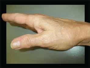
Figure 2: Shoulder sign.

Research suggests that compression caused by grip-and-pinch activities occurs over a relatively small surface area between the metacarpal and the trapezium and can lead to uneven wear patterns. Momose and colleagues examined the area of contact on the trapezium by the first metacarpal during various thumb movements.6 Their findings generally support what would be predicted by the convex/concave rules of joint motion. During opposition, the contact area on the trapezium was in the radial, volar, and ulnar segments. During abduction the contact area was in the dorsal segment.

ad goes here:advert-1
ADVERTISEMENT
SCROLL TO CONTINUE

Another notable finding of this study was the relatively small area of contact measured at the end ranges of movement. For opposition, the contact area measured approximately 50% of the articulating surfaces. For abduction, the contact area was approximately 25%. Microscopic analysis of cadaveric hands with thumb CMC joint OA identified that the location of greatest cartilage degeneration within the thumb CMC joint was in the dorsal radial region of the trapezium.7 One goal of therapy is to maintain maximum contact between articulating surfaces during joint motion to reduce compression within the joint. This can prevent uneven wear patterns in the thumb CMC joint and lessen the potential for cartilage deterioration. Knowledge of patients’ wear patterns can help physicians and therapists select management strategies for patients.

Muscle considerations: Forceful thumb use is required to hold objects between the thumb and fingers. As muscle force increases, so does the compression force between the trapezium and metacarpal. Pinching forces occur at the interface between the thumb and the fingertips in the case of a tip-to-tip or pad-to-pad pinch, and between the pad of the thumb and the lateral border of the index finger in the case of a lateral pinch. In either case, these forces occur approximately three to four inches from the axis of rotation at the thumb CMC joint. As described earlier, the muscles that generate forceful pinch cross the thumb CMC joint and act upon it. Pinching, an isometric activity, results in compression at the thumb CMC joint. Cooney calculated that the compression forces at the thumb CMC joint were approximately 12 times the pinching forces that occur at the thumb pad.8

ad goes here:advert-2
ADVERTISEMENT
SCROLL TO CONTINUE

Muscle imbalances between the finger muscles and thumb muscles also increase stress on the CMC joint. If the thumb muscles are weak in comparison to the finger muscles, the CMC joint will shift slightly into a position that increases point pressure within the joint during pinch. For example, during pad pinch the force of the finger muscles will produce thumb extension and abduction if unopposed by the thumb muscles. If the thumb muscles are incapable of resisting the force of the finger muscles, slight abduction and/or extension may occur, resulting in increased compression in the dorsal radial aspect of the trapezium. Beak ligament laxity will cause dorsal subluxation of the metacarpal, leading to volar surface compression on the trapezium. Opposition limited by weakness of the opponens, adductor tightness, or soft-tissue limitations of the web space will further exacerbate the problem. Figure 3a shows a pinching activity with good thumb and finger muscle balance while Figure 3b shows the same activity with limitations in thumb opposition. The stronger finger muscles, in addition to unstable ulnar collateral ligaments of the interphalangeal (IP) and MP joints, force the thumb into noticeable abduction at the IP and MP joints and more subtle abduction at the CMC joint. These positions result in point compression in the dorsal radial aspect of the trapezium.

Page: 1 2 3 4 5 6 7 | Single Page
Share: 

Filed under:ConditionsOsteoarthritis and Bone Disorders Tagged with:ExercisejointnonsurgicalOsteoarthritisPainpatient carerheumatologistTreatment

Related Articles
    Oksana Shufrych TKTK / Shutterstock.com

    Heated Gloves May Improve Hand Function in Diffuse Systemic Sclerosis

    October 16, 2017

    Systemic sclerosis (SSc), a subtype of scleroderma, is a rare, complex autoimmune disease characterized by widespread vasculopathy of the small arteries and fibroblast dysfunction.1,2 It has been described as a fibrosing micro­vascular disease, because vascular injury precedes and leads to tissue fibrosis.3 The resulting Raynaud’s phenomenon, pain, skin thickening and tightening, and multi-organ involvement have…

    ARHP News: Occupational and Hand Therapy in Treatment of Osteoarthritis

    November 2, 2012

    Osteoarthritis (OA) of the hand is one of the more frequent diagnoses seen in a hand clinic, though early referral to an occupational therapist can improve hand function in patients with OA

    Insights on the Diagnosis & Treatment of Low Back & Hip Pain

    March 19, 2019

    CHICAGO—Two experts presented insights on the diagnosis and treatment of low back and hip pain, including a refresher course on the mechanical structures involved, in Anatomy in a Day: Demystifying Low Back Pain and Lateral Hip Pain: New Patho-Anatomical Perspectives, a session at the 2018 ACR/ARHP Annual Meeting. Low Back Pain Avoid using such terms…

    New Tools for Myositis Diagnosis, Classification & Management

    April 15, 2019

    CHICAGO—At Hot Topics in Myositis, a session at the 2018 ACR/ARHP Annual Meeting, three experts discussed new classification criteria for idiopathic inflammatory myopathies (IIM) and offered practical primers on overlap myositis conditions and inclusion body myositis (IBM). New Myositis Classification Criteria After a 10-year development process, the new EULAR/ACR Classification Criteria for Adult and Juvenile…

  • About Us
  • Meet the Editors
  • Issue Archives
  • Contribute
  • Advertise
  • Contact Us
  • Copyright © 2025 by John Wiley & Sons, Inc. All rights reserved, including rights for text and data mining and training of artificial technologies or similar technologies. ISSN 1931-3268 (print). ISSN 1931-3209 (online).
  • DEI Statement
  • Privacy Policy
  • Terms of Use
  • Cookie Preferences