Given the foundation for the science of paleontology, your next question might be, “So what?”
Sorting Out Inflammatory Arthritis
Given the foundation for the science of paleontology, your next question might be, “So what?” Identifying the antiquity of a disease might interest a historian. Examining variation in its absence or presence and prevalence over time allows analysis of environmental interactions.4,11 For example, the frequency of spondylarthropathy is relatively low in humans, except when environmental (fecal–oral) contamination is present.11 This allows such contamination to be identified and, conversely, helped to refute some widely held theories such as the role of lead intoxication in Roman Italy. It now appears that use of lead coffins may have been responsible for the contamination of human remains from that era. After all, if saturnine gout from lead poisoning was rampant, wouldn’t gouty arthritis have been far more commonly observed among Romans?
Paleoepidemiology provides the opportunity to test current clinical perspectives of disease and the accuracy of our clinical diagnoses.12 It can help address whether disease categories are pure or whether they have been mistakenly coalesced into a single diagnosis.
A prime example of interest to rheumatologists is RA. Some of the confusion arises from the use of the term “RA” as a general appellation for peripheral joint inflammatory arthritis.4,12 This has been further confused by the term “rheumatoid spondylitis,” an old name for ankylosing spondylitis.13,14
Examination of the archeological record of North America reveals an intriguing window to the character of RA. The Western portions of the Tennessee and Green Rivers witnessed a disease manifest by a polyarticular erosive arthritis.15 Multiple populations with the disease shared common characteristics. Erosions were all marginally distributed and were not accompanied by reactive new bone or periosteal reaction, nor by joint fusion. Sacroiliac joints and vertebrae (C1–C2 excepted) were unaffected. Radiologic examination revealed periarticular osteopenia. Subchondral joint erosion and fusion of peripheral joints were not part of the disease. Thus, we seem to have multiple populations with only one disease present. This allowed an unfettered perspective of RA.
Examination of the zoologic and paleontologic record is even more instructive. While some researchers have observed isolated occurrences of “RA” in swine and dogs, examination of those cases reveals subchondral erosions, not only of peripheral joints, but also axial joint fusion, clearly a different disease than human RA.4,16-18 In fact, this disease is indistinguishable from human forms of spondylarthropathy. This condition has proven to be a pan-mammalian disorder, traceable as far back as the dinosaurs and beyond.19,20
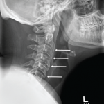
Similarly, paleopathology has allowed clarification of the character of other joint diseases. Calcium pyrophosphate deposition disease (CPPD) has been recognized by the radiographic findings of chondrocalcinosis and radiocarpal joint space narrowing.4,5 The latter characterizes wrist involvement, but is not amenable to study in unfleshed skeletons. However, joint space narrowing, perhaps sufficient for classification, was noted to be associated with clinical radiocarpal joint indentation that is recognizable in the skeletal record.21,22 Examination of skeletons with such radiocarpal indentation often revealed modification of the metacarpal phalangeal joints.1,4,21,22 The joint surfaces were incomplete and lacked the erosions of RA or the tophaceous lesions of gout. I have chosen the term “crumbled erosion” to describe this appearance.1,4,21 This observation not only provided a facile differential characteristic of erosions, but also permitted new insights into what had previously been called erosive osteoarthritis.23 Distal and proximal interphalangeal joints afflicted with this disease had the same epidemiologic features and radiologic changes characteristic of CPPD.21
Clues to Past Life
Paleopathology also provides clues to ancient lifestyles and habitats. For example, auropod dinosaurs (e.g., Apatosaurus, aka Brontosaur) were thought to spend substantial time in the water and climbing swamp or river banks. That would have put great stresses on the forefeet, but, paradoxically, stress fractures only affected their hind feet. Some researchers have speculated that they would stand on their hind feet, similar to ballet positions. However, the sauropods lacked the facet (zygapophyseal joint) and did not have the forefoot stress fractures characteristically noted in ballet dancers subject to such posture.24
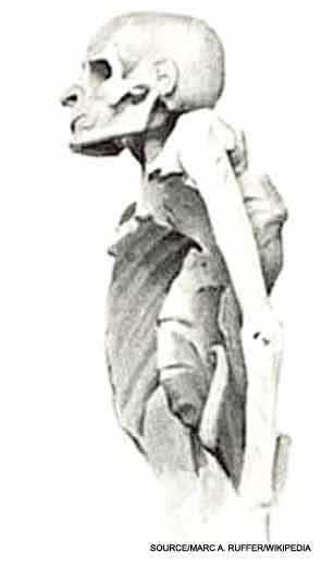
Perspectives of disease have, at times, been predicated upon historical accounts. These have to be analyzed to assure that ancient diagnoses actually correlate with modern diagnoses. The written record must be analyzed from primary sources. Leprosy is the classic example of a disease that has been misunderstood by reading the ancient records. Studies of archeological “leprosy cemeteries” created a perspective of disease that could not be correlated with what was found in contemporarily diagnosed populations.25 The archeological sites were “leprosy cemeteries” related to the Leprosauria that Pope Clement closed in 1508 and 1510. There were approximately 10,000 cemeteries, each housing at least 100 bodies. This suggests that more than a million individuals were afflicted with leprosy, a number that seems illogical. So, what did leprosauria actually represent, and who was buried in their associated cemeteries? Examination of diagnostic categorization in the 15th and 16th centuries reveals that many skin disorders were classified as/with leprosy. For example, psoriatic and reactive arthritis have associated joint destruction with cutaneous changes that could have been confused with leprosy. Most of the individuals in these cemeteries have skeletal changes that are now recognized as being characteristic of the spondylarthropathies.25
The foundation for scientific paleopathology is the establishment of sufficient skills and experience to accurately identify physical findings. Accuracy replaces the previous precision-based approach, where precision is defined as adherence to authority-based perspectives, which vary among those who claim to be authorities.4,26 The latter is compromised because critical scientific methodology was apparently not understood.
The classic exemplar is the challenge that periosteal reaction presents to the field of anthropology.26 Remodeling of the outer layer of cortical bone can have many etiologies, ranging from healing trauma to infection to cancer. Trauma may directly alter bone surfaces (e.g., bone bruises) or may manifest as a cumulative effect (e.g., stress fracture). Infectious damage may be related to pyogenic, tuberculous, fungal, or treponemal disease.1,4 Among anthropologists, there was a view that most occurrences of periosteal reaction in populations must represent the result of trauma.27 That speculation does not withstand scrutiny. No correlation was noted in populations with significant environmental hazards.28,29 As an example, periosteal reaction was absent in hostile environments such as the Northwest Territories of Canada, but was common in the Nubian desert.
Beyond this speculation, an even more fundamental error has been the lack of validation of the investigators’ skill in recognizing periosteal changes.26 The skeletons from the Irene Mound site in Georgia have been the source for many publications. Limiting the analysis of the investigators skills to the tibiae, the results obtained have identified two camps. One group perceived periosteal reaction to be present in 100% of skeletons, while the other group described the complete absence of this finding. My team’s examination achieved an intermediate result. Who is correct? Accuracy can only be claimed with independent verification. Independent verification not only requires selection of the appropriate test, but also that the results be interpreted in light of understanding of disease pathophysiology and/or its transmission through ensuing generations.
This approach can be understood with examination of a phenomenon recognized in Egyptian mummies.30 Standard X-rays suggested calcification of intervertebral discs. The presence of associated black pigmentation suggested that an inherited disease of metabolism, ochronosis, was common among Egyptians. In an effort to independently validate this diagnosis, infrared spectroscopy was used to confirm that the deposition product was homogentisic acid. This compound was noted in half of the Egyptian mummies. When one considers that the transmission pattern of ochronosis is via recessive genes, there is a dilemma. This genetic pattern would predict that no more than one quarter of the population should be afflicted.31 How do we explain this paradox? While routine X-rays suggested generalized disc calcification, CT revealed that the “calcification” was misread and was the result of overlapping shadows caused by multiple calcific nodules. Nuclear magnetic resonance spectroscopy demonstrated that the compound was not homogentisic acid. It identified the embalming material used by ancient Egyptians and that the nodules were salt natrons derived from the same process.
Returning to the validation of the causes of periosteal bone reactions, physical chemistry came to the rescue. After raising bone temperature by three degrees above ambient temperature, the time course of the tissues’ return to the original ambient temperature was assessed.26 It was suspected that the previous recognition of periosteal reaction had been compromised by the inability to distinguish periosteal reaction from a postmortem taphonomic process, bone abrasion, which exposed layers of intracortical bone. Periosteal reaction occurs on top of the original cortical surface, while abrasion exposes the subsurface bone. The time course of heat dissipation differed between abraded bone and that caused byperiosteal reaction, with no overlap in the time course. Taphonomically altered bone revealed a prolonged heat dissipation time compared to bones remodeled by periosteal reaction. Recently, epi-illumination microscopy has been shown to be a useful technique in demonstrating the shaved appearance of abraded bone in contrast to the prominent surface vascular channels seen with periosteal reaction bone remodeling.
Paleopathology has developed from a speculative endeavor—related predominantly to curiosities—to a science, which contributes to our understanding of the past. It can provide insights to improve the future health for the denizens of earth, both animal and vegetal, often despite human interferences.
Dr. Rothschild is professor of medicine at Northeast Ohio Medical University in Rootstown, and in the departments of geology and anthropology at the University of Kansas in Lawrence.
References
- Rothschild BM, Schultze H-P, Peligrini R. Herpetological Osteopathology: Annotated Bibliography of Amphibians and Reptiles. Heidelberg, Germany: Springer-Verlag, 2013.
- Ehrlich GE. Osteoarthritis beginning with inflammation: Definitions and correlations. JAMA. 1975;232:157-159.
- Waldron HA. Prevalence and distribution of osteoarthritis in a population from Georgian and early Victorian London. Ann Rheum Dis. 1991;50:301-307.
- Rothschild BM, Martin LD. Skeletal Impact of Disease. New Mexico Museum of Natural History, 2006.
- Resnick D. Radiology of Bone and Joint Disease. Philadelphia: Saunders, 2002.
- Hacking P, Allen T, Rogers, J. Rheumatoid arthritis in a medieval skeleton. Intl J Osteoarcheol. 1994;4:251-255.
- Rogers J, Waldron T, Dieppe P, Watt, I. Arthropathies in palaeopathology: The basis of classification according to most probable cause. J Archaeol Sci. 1987;14:179-193.
- Ortner DJ, Putschar W. Identification of Paleopathological Conditions in Human Skeletal Remains. Smithsonian Institution Press: Washington, DC, 1985.
- Rothschild BM, Woods RJ, Ortel W. Rheumatoid arthritis “In the buff”: Erosive arthritis in representative defleshed bones. Amer J Phys Anthropol. 1990;82:441-449.
- Rothschild BM, Woods RJ. Spondyloarthropathy: Erosive arthritis in representative defleshed bones. Am J Phys Anthropol. 1991;85:125-134.
- Rothschild BM, Rothschild C. Nineteenth century spondyloarthropathy independent of socioeconomic status: Lack of skeletal collection bias. J Rheumatol. 1993;20:314-319.
- Rothschild BM. Two faces of “rheumatoid arthritis”: Type A versus type B disease. J Clin Rheumatol. 1997;3:334-338.
- Aufderheide AC, Rodriguez-Martin C. The Cambridge Encyclopedia of Human Paleopathology. Cambridge University Press: Cambridge, U.K., 1998.
- Present AJ. Rheumatoid spondylitis. Cal West Med. 1945;63:10-13.
- Rothschild BM, Woods RJ, Rothschild C, Sebes JI. Geographic distribution of rheumatoid arthritis in ancient North America: Implications for pathogenesis. Semin Arthritis Rheum. 1992;22:181-187.
- Carr AP, Michels G. Identifying noninfectious erosive arthritis in dogs and cats. Vet Med. 1997;92:804-810.
- Sikes D. A rheumatoid-like arthritis in swine. Lab Invest. 1959;8:1406-1415.
- Rothschild BM, Rothschild C, Woods JR. Inflammatory arthritis in canids: Spondyloarthropathy. J Zoo Wildlife Med. 2001;32:58-64.
- Rothschild BM, Rothschild C. Spondyloarthropathy mammalian as a trans-phenomenon, reproducible in its manifestations across species lines. J Paleopath. 2000;11:103-104.
- Rothschild BM. Radiologic assessment of osteoarthritis in dinosaurs. Ann Carnegie Museum. 1990;59:295-301.
- Rothschild BM, Woods RJ, Rothschild C. Calcium pyrophosphate deposition disease: Description in defleshed skeletons. Clin Exp Rheumatol. 1992;10:557-564.
- Rothschild BM, Yakubov LE. Prospective six month, double-blind trial of hydroxychloroquine treatment of calcium pyrophosphate deposition disease. Contemp Ther. 1997;23:327-331.
- Peter JB, Pearson CM, Marmor L. Erosive osteoarthritis of the hands. Arthritis Rheum. l966: 9:365-368.
- Rothschild BM, Molnar RE. Sauropod stress fractures as clues to activity. In: Thunder-lizards. Tidwell V, Carpenter K, eds. Bloomington: Indiana University Press; 2005:381-391.
- Rothschild BM, Rothschild C. Skeletal manifestations of leprosy: Analysis of 137 patients from different clinical settings in the pre- and post-modern treatment eras. J Clin Rheumatol. 2001;7:228-237.
- Rothschild BM, Rothschild C. Thermodynamic resolution of periosteal reaction and taphonomic change. Reumatismo. 2003;55:195-201.
- Heathcote GM, Stodder AL, Buckley HR, et al. Treponemal disease in the Western Pacific: Corrections and critique. Curr Anthropol. 1998;40:359-368.
- Rothschild C, Rothschild BM. Occurrence and transitions among the treponematoses in North America. Chungara, Revista de Antropologia Chilena. 2000;32:147-155.
- Rothschild BM, Rothschild C. Analysis of treponemal disease in North Africa: The case for Bejel in the Sudan, but absence in West North Africa. Hum Evol. 1996;11:11-15.
- Stenn FF, Milgram JW, Lee SL, Weigand RJ, Veis A. Biochemical identification of homogentisic acid pigment in an ochronotic Egyptian mummy. Science. 1977;197:566-568.
- Stanbury JB, Wyngaarden JB, Fredrickson DS, Goldstein JL, Brown MS. The Metabolic Basis of Inherited Disease. 5th ed. New York, McGraw Hill, 1983.