Video: Every Case Tells a Story| Webinar: ACR/CHEST ILD Guidelines in Practice

An official publication of the ACR and the ARP serving rheumatologists and rheumatology professionals

  • Conditions
    • Axial Spondyloarthritis
    • Gout and Crystalline Arthritis
    • Myositis
    • Osteoarthritis and Bone Disorders
    • Pain Syndromes
    • Pediatric Conditions
    • Psoriatic Arthritis
    • Rheumatoid Arthritis
    • Sjögren’s Disease
    • Systemic Lupus Erythematosus
    • Systemic Sclerosis
    • Vasculitis
    • Other Rheumatic Conditions
  • FocusRheum
    • ANCA-Associated Vasculitis
    • Axial Spondyloarthritis
    • Gout
    • Psoriatic Arthritis
    • Rheumatoid Arthritis
    • Systemic Lupus Erythematosus
  • Guidance
    • Clinical Criteria/Guidelines
    • Ethics
    • Legal Updates
    • Legislation & Advocacy
    • Meeting Reports
      • ACR Convergence
      • Other ACR meetings
      • EULAR/Other
    • Research Rheum
  • Drug Updates
    • Analgesics
    • Biologics/DMARDs
  • Practice Support
    • Billing/Coding
    • EMRs
    • Facility
    • Insurance
    • QA/QI
    • Technology
    • Workforce
  • Opinion
    • Patient Perspective
    • Profiles
    • Rheuminations
      • Video
    • Speak Out Rheum
  • Career
    • ACR ExamRheum
    • Awards
    • Career Development
  • ACR
    • ACR Home
    • ACR Convergence
    • ACR Guidelines
    • Journals
      • ACR Open Rheumatology
      • Arthritis & Rheumatology
      • Arthritis Care & Research
    • From the College
    • Events/CME
    • President’s Perspective
  • Search

What Rheumatologists Should Look for in Diagnosing SAPHO Syndrome

Richard Brasington, MD  |  Issue: November 2012  |  November 1, 2012

Skin Lesions in SAPHO Syndrome
click for large version
Table 1: Skin Lesions in SAPHO Syndrome

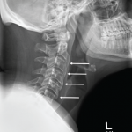
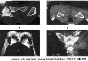
Osteoarticular involvement is especially common in the anterior chest wall, axial skeleton, and large joints such as the wrists, knees, hips, and ankles. The clavicles, sternum, and sterno-clavicular joints are often involved as well (see Figure 5). Spondylodiscitis and unilateral sacroiliitis may be seen on imaging of the axial skeleton.

ad goes here:advert-1
ADVERTISEMENT
SCROLL TO CONTINUE

A major diagnostic dilemma often occurs because the early bone lesions of SAPHO are indistinguishable from those seen in cases of infectious osteomyelitis. In addition, the inflammatory synovial fluid and the destructive lesions seen on imaging require the clinician to seriously consider a septic arthritis and/or osteomyelitis in the differential diagnosis. With time, there may be neutrophilic infiltration with pseudoabscess formation. Ultimately, the bony trabeculae become sclerotic and enlarged. The finding of a “culture negative osteomyelitis” should prompt the rheumatologist to consider the diagnosis of SAPHO.

ad goes here:advert-2
ADVERTISEMENT
SCROLL TO CONTINUE
Computed tomography scans of A, the anterior chest wall showing sternocostoclavicular hyperostosis with subchondral erosions (arrows), B, soft tissue thickening and calcifications around the medial head of the clavicle (arrow) in a 62-year-old man with a disease duration of 6.9 years, C, hyperostosis and grotesque periostitic lesions, and D, massive osteitis with diffuse and osteolytic areas.
Figure 5: Computed tomography scans of A, the anterior chest wall showing sternocostoclavicular hyperostosis with subchondral erosions (arrows), B, soft tissue thickening and calcifications around the medial head of the clavicle (arrow) in a 62-year-old man with a disease duration of 6.9 years, C, hyperostosis and grotesque periostitic lesions, and D, massive osteitis with diffuse and osteolytic areas.

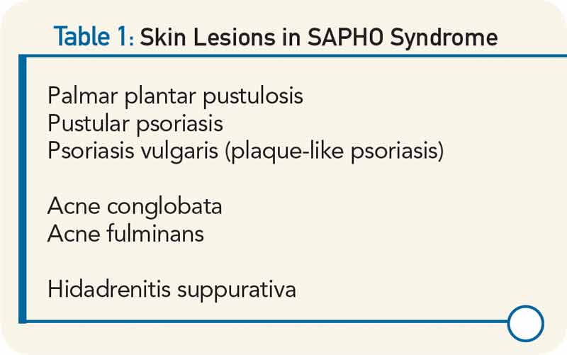
A series of 71 patients has been described by Colina et al.1 Forty-eight of the 71 patients (68%) were female. Eighty percent had skin disease. The mean age of onset was 38.5 years, but the mean age of diagnosis was 45.5 years. Seventy percent of patients had anterior chest wall involvement, 24% described inflammatory back pain, and only 11% had a peripheral joint synovitis. Fifty-six percent had some form of skin involvement, most commonly PPP.

Markers of inflammation were uncommonly elevated. Only 21% of patients had an elevated erythrocyte sedimentation rate, and only 17% demonstrated elevated C-reactive protein. In this series, only 4% of patients were human leukocyte antigen B-27 antigen positive.

Examining Causes

The pathophysiology of SAPHO remains obscure. Many investigators have speculated that there is a bacterial cause for this condition. For example, in one German study, 14 of 21 (67%) needle biopsy specimens of osteitis lesions were culture positive for P. acnes. These 14 patients were combined with 16 other patients who did not have bone cultures performed. All were treated with azithromycin for 16 weeks, chosen because it is concentrated in phagocytic cells and because macrolide antibiotics may have antiinflammatory activity. After 16 weeks of therapy, improvement was noted in the skin, osteitis, and MRI scores. However, when azithromycin treatment was withdrawn after 28 weeks of therapy, the previously noted improvement had regressed.

Page: 1 2 3 4 5 | Single Page
Share: 

Filed under:Clinical Criteria/GuidelinesConditionsOsteoarthritis and Bone Disorders Tagged with:anti-inflammatorybonecase reportimaginginfliximabPainpatient careradiographrheumatologistSAPHOsynovitisTreatment

Related Articles

    Case Report: A Patient Presents with Rare, Fulminant SAPHO Syndrome

    October 18, 2018

    Synovitis, acne, pustulosis, hyperostosis and osteitis (SAPHO) syndrome is a heterogeneous, inflammatory, musculoskeletal disease. The disease is an insidious, sterile osteitis with associated skin and synovial inflammation.1 Diagnosis can prove challenging, but a thorough clinical history, high clinical suspicion and imaging techniques can help clinch it. The below case reveals a rare, fulminant presentation of…

    Case Report: Acne Drug Leads Patient to Retinoid Hyperostosis

    November 19, 2018

    Presentation A 26-year-old man with a history of acne vulgaris and hidradenitis suppurativa presented to our rheumatology clinic with persistent back pain and stiffness of three years’ duration. He described bilateral low back pain that was worse when he arose in the morning and at night when he was trying to sleep. In a similar…

    Diagnostic Drill-Down: New Research Suggests Genetic Autoinflammatory Roots

    January 25, 2021

    Two experts discussed ongoing difficulties in diagnosing autoinflammatory disease, & promising new studies that point to possible genetic roots of autoinflammatory disease.

    Imaging in Ankylosing Spondylitis

    April 1, 2015

    MRI inflammation, fat and new bone formation in the sacroiliac joints, spine in patients with AS

  • About Us
  • Meet the Editors
  • Issue Archives
  • Contribute
  • Advertise
  • Contact Us
  • Copyright © 2025 by John Wiley & Sons, Inc. All rights reserved, including rights for text and data mining and training of artificial technologies or similar technologies. ISSN 1931-3268 (print). ISSN 1931-3209 (online).
  • DEI Statement
  • Privacy Policy
  • Terms of Use
  • Cookie Preferences