The American College of Rheumatology (ACR) has launched the long-awaited certification program for musculoskeletal (MSUS) ultrasound in rheumatology
Search results for: musculoskeletal ultrasound
ACR Musculoskeletal Ultrasound Education
The ACR created the Musculoskeletal Ultrasound Course for Rheumatologists with Interventional Cadaver Workshop—Intermediate specifically for rheumatologists who are using musculoskeletal ultrasound in their practice and are ready to expand their skills.
New Musculoskeletal Ultrasound Course
The ACR is broadening its educational offerings in musculoskeletal ultrasound by holding its first stand-alone musculoskeletal ultrasound course for rheumatologists this August in Chicago. The same course will be offered prior to the 2010 ACR/ARHP Annual Scientific Meeting.
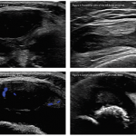
The Role Ultrasound Imaging Plays in Diagnosing Hemangiomas
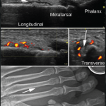
A 17-year-old woman presents with chronic finger pain experienced over six months that is worse in the mornings. On physical exam, the patient has no joint swelling, pain on range of motion or limitation of range of motion in any of her finger joints. She has a tender, subcutaneous, firm, flesh-colored nodule on the lateral…
The Power of Power Doppler: Ultrasound Imaging in RA
At the 17th Annual Advances in the Diagnosis & Treatment of the Rheumatic Diseases meeting, Dana DiRenzo, MD, MHS, RhMSUS, discussed the use of ultrasound imaging in patients with inflammatory arthritis.
Ultrasound Provides Insights into Immune Checkpoint Inhibitor‐Induced Inflammatory Arthritis
Ultrasound may provide unique insights into the effects of immune checkpoint inhibitors on the human body beyond the immune system. Research suggests synovitis and inflammatory tendon involvement are commonly seen in patients with immune checkpoint inhibitor-induced inflammatory arthritis.
Ultrasound in Rheumatology—Past, Present & Future
For most rheumatologists, the key elements of the physical exam—inspection, palpation, percussion and auscultation—have long been second nature, but a fifth modality has grown in importance with respect to making the correct diagnosis: ultrasound. From evaluating for Doppler signal and additional findings indicative of synovitis to identifying bony erosions, chondrocalcinosis, tophi and other articular and…

Tips for Implementing Ultrasound Training in Rheumatology Fellowships
ATLANTA—Point-of-care ultrasound education mainly has occurred at the undergraduate level at U.S. medical schools, but rheumatology fellowship training programs are rapidly catching up and integrating it into their curricula, according to two program directors who reviewed the state of rheumatology ultrasound education, including potential barriers to its implementation, on Nov. 12 at the 2019 ACR/ARP…
Ultrasound Image Review: A 30-Year-Old Woman with Left Foot Pain
Presentation A 30-year-old woman presented to her rheumatologist for left foot pain of three weeks’ duration. She was followed for systemic lupus erythematosus manifesting in arthritis and hemolytic anemia, as well as anti-nuclear antibody and Smith antibody positivity, and was treated with hydroxychloroquine and prednisone in the 2.5–10 mg per day range. She was symptom…
The Diagnostic View: Ultrasound of a Child’s Sore Knee
Editor’s note: In this recurring feature, we first present a series of ultrasound images for your review, and then a brief discussion of the findings and diagnosis. Before you scroll to the discussion, examine these images carefully and draw your own conclusions. History A 2-year-old boy with a history of multiple strokes and vertebral artery…
- « Previous Page
- 1
- 2
- 3
- 4
- …
- 20
- Next Page »