Highlights from the 2017 EULAR Congress MADRID—Researchers say that whole-body MRI could yield an earlier diagnosis of spondyloarthropathy (SpA) in patients with early inflammatory joint symptoms, according to findings presented in a poster session at the Annual European Congress of Rheumatology (EULAR). The approach could lead to earlier treatment and better outcomes, they say. Investigators…
Ultrasound Can be Useful in Diagnosing Gout
The presence of synovial monosodium urate monohydrate (MSU) crystals is the gold standard for diagnosing gout. But a new study, funded in part by the ACR and led by rheumatologists, including Alexis Ogdie, MD, MSCE, evaluated the effectiveness of ultrasound in diagnosing it. The study found that ultrasound can be useful in discriminating gout from non-gout….
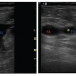
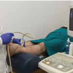
Rheumatology Case Report: Deep Vein Thrombosis Detected by Point-of-Care Ultrasound
Case A 46-year-old Caucasian female presented to the outpatient rheumatology clinic where she had been followed for several years. Her chief complaint was pain in her right knee, posterior right thigh and right hip that had begun gradually over the previous three weeks. Her past medical history was significant for rheumatoid arthritis (RA), obesity and…
fMRI Can Help Diagnose Fibromyalgia
Brain imaging can distinguish fibromyalgia patients from healthy controls with high sensitivity and specificity, according to two papers published nearly simultaneously in Pain late last summer, by groups at the Universities of Colorado and Michigan, respectively. Somewhat surprisingly to the authors and others, in the Colorado study, which used both painful and nonpainful stimuli, the…
Ultrasound May Be Useful for Grading Rotator Cuff Tendinopathy
Researchers have developed procedures and assessed their efficacy for the use of ultrasound images to measure the inter-rater reliability of the measurement of structural changes in the tendon of patients with supraspinatus tendinopathy. The standardized procedures proved useful in evaluating patients…
Diagnostic Imaging in Patient with Chronic Left Ankle Pain: History
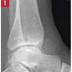
Editor’s note: In this recurring feature, we first present a series of images (this page) for your review, and then a brief discussion of the findings and diagnosis. Before you turn to the discussion, examine these images carefully and draw your own conclusions. History A 49-year-old woman presents with one year of chronic left ankle…
Diagnostic Imaging in Patient with Chronic Left Ankle Pain: Findings
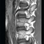
Radiographic imaging showed circumferential soft tissue swelling of the ankle with a soft-tissue density seen in the tibiotalar and posterior subtalar joints, as well as a large, lobulated effusion. MRI of the left ankle shows cystic changes within the talus and first cuneiform bones, as well as a lobulated abnormal soft tissue density with low…
Diagnostic Imaging in Lupus Patient with Foot Pain: Findings
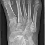
View the question. Findings/Diagnosis An anteroposterior (AP) radiograph of the right foot shows hallux valgus of the first metatarsal phalangeal (MTP) joint, erosive changes at the first and fifth metatarsal bones and degenerative changes at the fourth and fifth metatarsal-cuboid joints. An AP radiograph of the left foot shows extensive erosive and degenerative changes at…

Diagnostic Imaging in Lupus Patient with Foot Pain: History
Editor’s note: In this recurring feature, we first present a series of images (this page) for your review, and then a brief discussion of the findings and diagnosis. Before you turn to the discussion, examine these images carefully and draw your own conclusions. History A 33-year-old woman with a 16-year history of systemic lupus erythematosus…
EULAR 2015: Imaging in Rheumatology
ROME, Italy—The explosion of imaging technology has made it more important than ever to establish a standardized way in which imaging can and should be used in clinical practice, an expert said in a session at EULAR 2015, the annual congress of the European League Against Rheumatism (EULAR). Marie-Antonietta d’Agostino, MD, PhD, professor of rheumatology…
- « Previous Page
- 1
- 2
- 3
- 4
- 5
- 6
- …
- 13
- Next Page »