Video: Every Case Tells a Story| Webinar: ACR/CHEST ILD Guidelines in Practice

An official publication of the ACR and the ARP serving rheumatologists and rheumatology professionals

  • Conditions
    • Axial Spondyloarthritis
    • Gout and Crystalline Arthritis
    • Myositis
    • Osteoarthritis and Bone Disorders
    • Pain Syndromes
    • Pediatric Conditions
    • Psoriatic Arthritis
    • Rheumatoid Arthritis
    • Sjögren’s Disease
    • Systemic Lupus Erythematosus
    • Systemic Sclerosis
    • Vasculitis
    • Other Rheumatic Conditions
  • FocusRheum
    • ANCA-Associated Vasculitis
    • Axial Spondyloarthritis
    • Gout
    • Psoriatic Arthritis
    • Rheumatoid Arthritis
    • Systemic Lupus Erythematosus
  • Guidance
    • Clinical Criteria/Guidelines
    • Ethics
    • Legal Updates
    • Legislation & Advocacy
    • Meeting Reports
      • ACR Convergence
      • Other ACR meetings
      • EULAR/Other
    • Research Rheum
  • Drug Updates
    • Analgesics
    • Biologics/DMARDs
  • Practice Support
    • Billing/Coding
    • EMRs
    • Facility
    • Insurance
    • QA/QI
    • Technology
    • Workforce
  • Opinion
    • Patient Perspective
    • Profiles
    • Rheuminations
      • Video
    • Speak Out Rheum
  • Career
    • ACR ExamRheum
    • Awards
    • Career Development
  • ACR
    • ACR Home
    • ACR Convergence
    • ACR Guidelines
    • Journals
      • ACR Open Rheumatology
      • Arthritis & Rheumatology
      • Arthritis Care & Research
    • From the College
    • Events/CME
    • President’s Perspective
  • Search

Fellows’ Forum Case Report: Tophaceous Bullae

Mark Vercel, DO, Kim Reinhart, OMS-3, & Amita Thakkar, MD  |  Issue: August 2017  |  August 13, 2017

As a manifestation of hyperuricemia, inflammatory bullous lesions have rarely been described in the past century. A more classic presentation of hyperuricemia is acute inflammatory gouty arthritis, characterized by the deposition of monosodium urate crystals. Other complications of chronic untreated hyperuricemia may include polyarticular arthritis, tophus formation and possible chronic destructive lesions of the bone, leading to permanent disability. Cutaneous tophus formation is one possible manifestation of chronic hyperuricemia and can be associated with acute inflammatory changes, such as a gouty cellulitis and, rarely, bullous lesions.

Case

An 87-year-old woman with a history of untreated polyarticular gout was admitted to the hospital due to acute pain in several joints, including both hands and her right foot, with spontaneous formation of new blister-like lesions on the hands. Her past medical history included treatment with torsemide for congestive heart failure from ischemic cardiomyopathy, coronary artery disease, type II diabetes mellitus, hyperlipidemia, hypertension, atrial fibrillation with embolic stroke and chronic normocytic anemia. She also had a history of previously untreated gouty episodes occurring over the previous eight years.

Figures 1A & 1B

Figures 1A & 1B
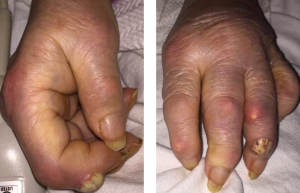
Fig. 1A: Creamy, yellow-hued bullae present over the DIP of the second digit of the right hand; and Fig. 1B: DIP of the fourth digit of the left hand with associated polyarticular synovitis. Tophi are seen overlying the PIP of the second digit.

Upon presentation to the hospital, she was afebrile with normal vital signs. She had podagra of the right great toe, and synovitis of the MCPs of multiple joints of the hands, in an asymmetric fashion. She demonstrated multiple cutaneous tophi, as well as multiple warm, tender, erythematous tophaceous lesions overlying the distal interphalangeal (DIP) and proximal interphalangeal (PIP) joints. The patient confirmed that these tophi are typically not inflamed and have been present for several years. However, she had two new lesions overlying the right second DIP and left fourth DIP, which were exquisitely tender, and had a tense, bullous appearance and texture with a creamy, yellow-white discoloration overlying an erythematous base (see Figures 1A and B).

She had a white blood cell count of 22,200/µL, hemoglobin of 10.5 g/dL, platelet count of 688,000/µL. Her renal function was within normal limits, and her serum uric acid level was 12.5 mg/dL. Radiographic examination of the feet and hands (see Figure 2) showed multiple destructive, erosive lesions with overhanging edges, consistent with a chronic, erosive, gouty arthritis.

ad goes here:advert-2
ADVERTISEMENT
SCROLL TO CONTINUE

Page: 1 2 3 4 5 | Single Page
Share: 

Filed under:Conditions Tagged with:case reportClinicalFellowsFellows ForumoutcomeRheumatic DiseaserheumatologyTophaceous Bullae

Related Articles

    Clinical Applications of Dual-Energy Computed Tomography for Rheumatology

    June 1, 2014

    Advanced imaging technique allows physicians to detect deposition of monosodium urate crystals not apparent in physical exams and better diagnose gout

    Difficult Gout

    July 1, 2007

    “Grandpapa’s Torments” was the Rodnan Commemorative Gout Print featured at the 2005 ACR/ARHP Annual Scientific Meeting.

    Lost and found

    Understanding the Role of Uric Acid in Gout

    September 6, 2022

    From the first substantial argument in the 19th century that uric acid played a role in gout, it took about 100 years for the medical community to accept its role in triggering acute inflammatory gout attacks. Two papers, both published in 1962, helped demonstrate the link between uric acid and acute gout attacks, quickly opening…

    Gout, Glucose Metabolism and Obesity: A Case Review

    November 2, 2014

    New research explores association between hyperurecimia and gout with metabolic derangement

  • About Us
  • Meet the Editors
  • Issue Archives
  • Contribute
  • Advertise
  • Contact Us
  • Copyright © 2025 by John Wiley & Sons, Inc. All rights reserved, including rights for text and data mining and training of artificial technologies or similar technologies. ISSN 1931-3268 (print). ISSN 1931-3209 (online).
  • DEI Statement
  • Privacy Policy
  • Terms of Use
  • Cookie Preferences